What is your diagnosis of this renal transplant patient?
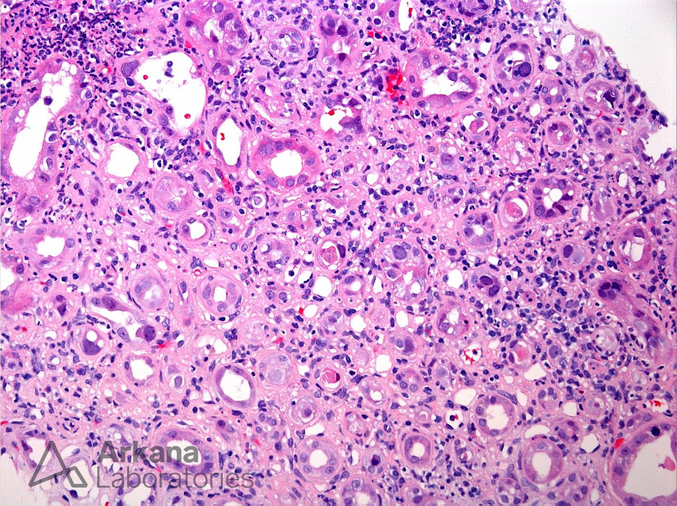
This H&E photomicrograph shows interstitial inflammation and tubular atrophy. Within tubules, multiple epithelial nuclei show irregularity such as enlargement, intranuclear inclusions, and dark staining (heterochromasia). These findings are consistent with viral nuclear changes but not specific for a particular viral etiology. However, in the setting of renal transplant these findings likely represent BK/Polyoma nephritis as it is markedly more common than other potential viral etiologies (CMV and adenovirus) and the morphology better fits with this etiology. An SV40 stain was performed (not shown) and showed positivity within numerous tubular nuclei throughout the sample confirming BK/Polyoma virus nephritis.
Quick note: This post is to be used for informational purposes only and does not constitute medical or health advice. Each person should consult their own doctor with respect to matters referenced. Arkana Laboratories assumes no liability for actions taken in reliance upon the information contained herein.