What’s your diagnosis?
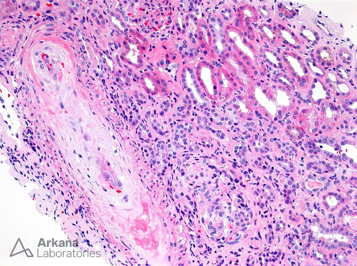
This photomicrograph shows an intrarenal artery with mucoid intimal expansion and near complete lumen obstruction. Within the expanded intima we see occasional RBCs and toward the center we see markedly reactive endothelial cells. These findings are consistent with a thrombotic microangiopathy (TMA). In this case, the TMA predominantly involved the vessels with most glomeruli showing only ischemic changes. While all forms of TMA have overlapping morphology making them difficult to segregate on biopsy, this type of vascular predominant TMA is often seen in the setting of accelerated/malignant hypertension, scleroderma renal crisis, drug use and sometimes antiphospholipid antibody syndrome. This patient had a history of illicit Opana use via IV injection which has been associated with TMA and this morphology. Please see the link for further reading on Opana-associated TMA.
Quick note: This post is to be used for informational purposes only and does not constitute medical or health advice. Each person should consult their own doctor with respect to matters referenced. Arkana Laboratories assumes no liability for actions taken in reliance upon the information contained herein.