A 24 day-old phenotypic female baby presents with nephrotic syndrome. Her medical history is significant for severe combined immunodeficiency syndrome (SCIDS) (diagnosed during newborn screen) and congenital nephrotic syndrome. She did not have any dysmorphic features. There is no history of premature delivery or other birth defects. Parents are of Native American ancestry. Investigations show serum albumin of 1.2 g/dL. Urine protein/creatinine ratio is 44. Investigations for TORCH infections are negative. Serological studies including HIV are also negative.
The images are characteristic findings of which of the following?
1. Diffuse mesangial sclerosis
2. Denys-Drash Syndrome
3. Minimal change disease
4. Congenital nephrotic syndrome of the Finnish type
5. Alport syndrome
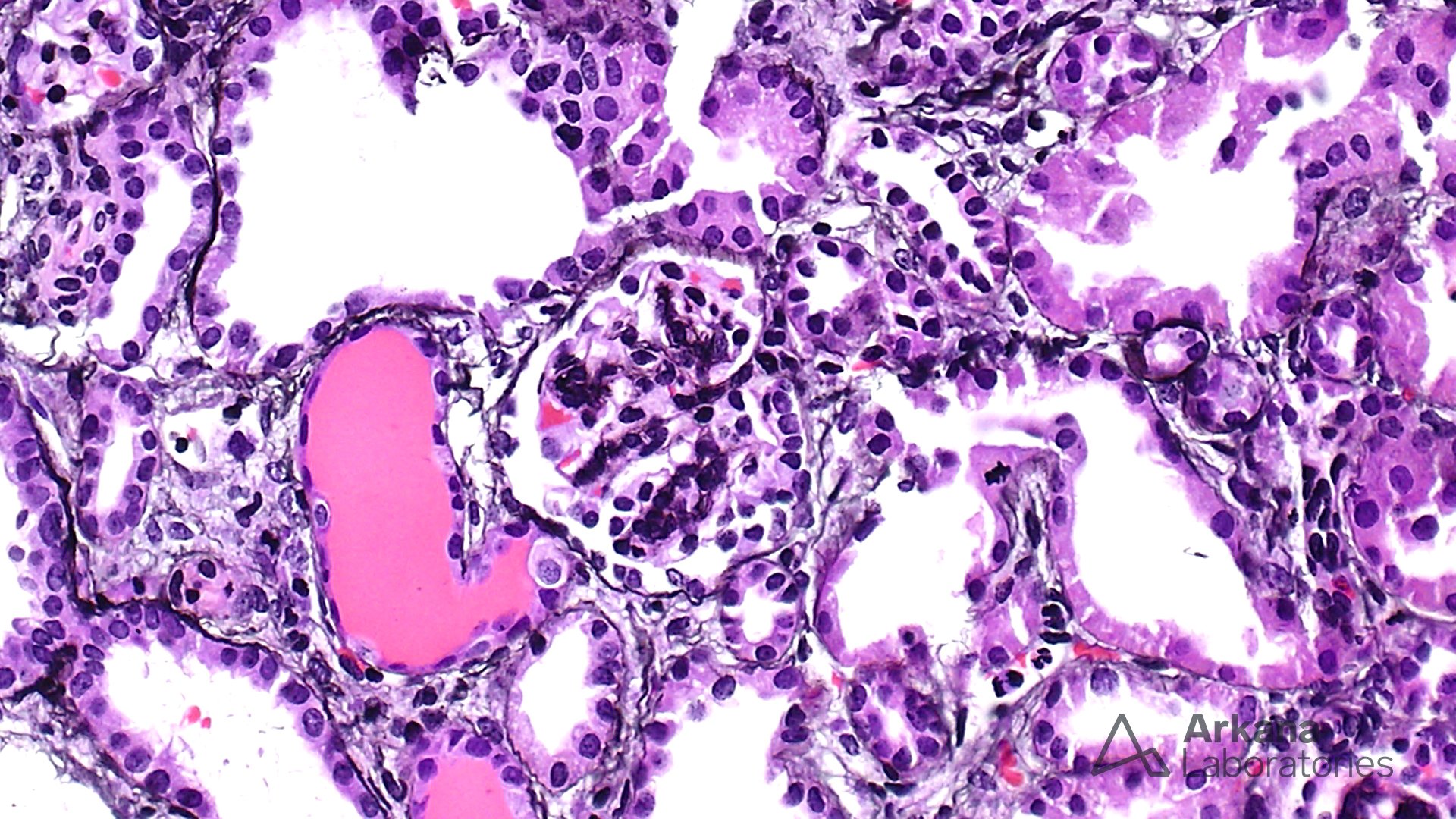
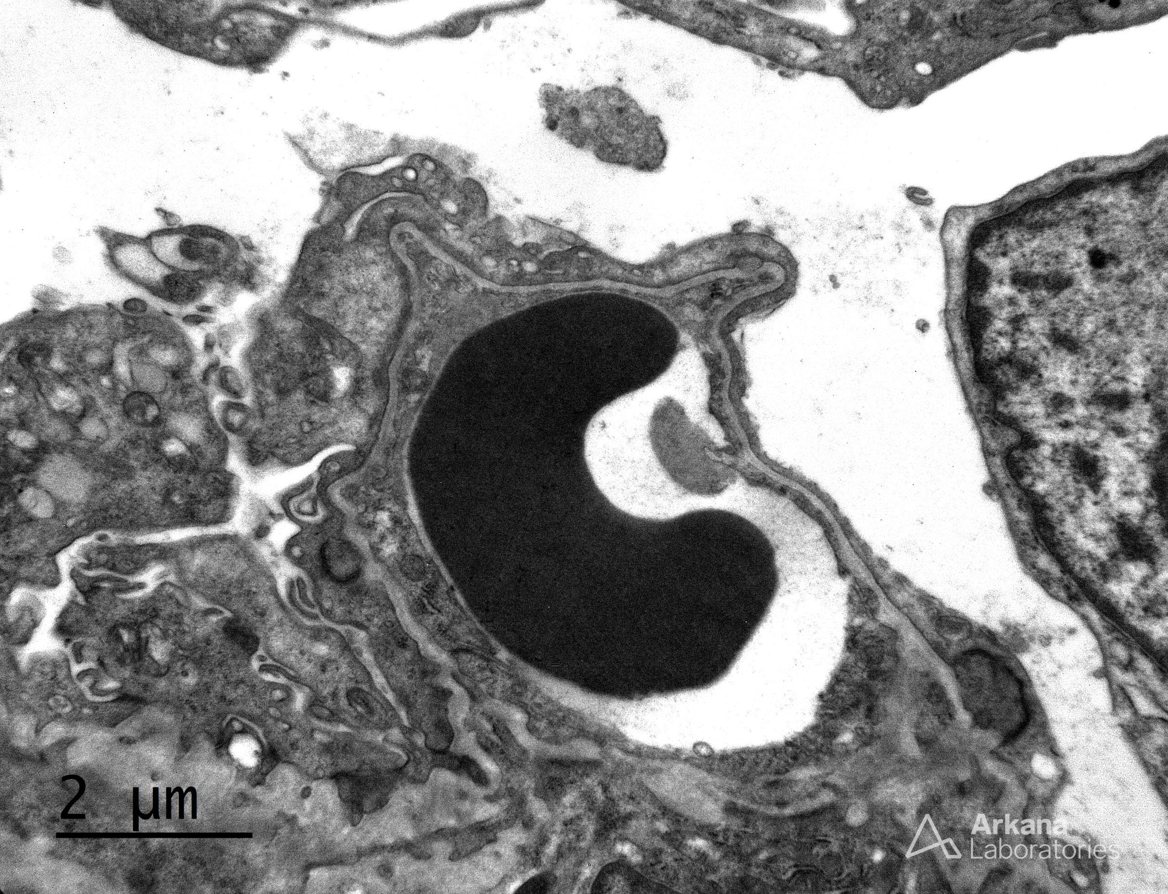
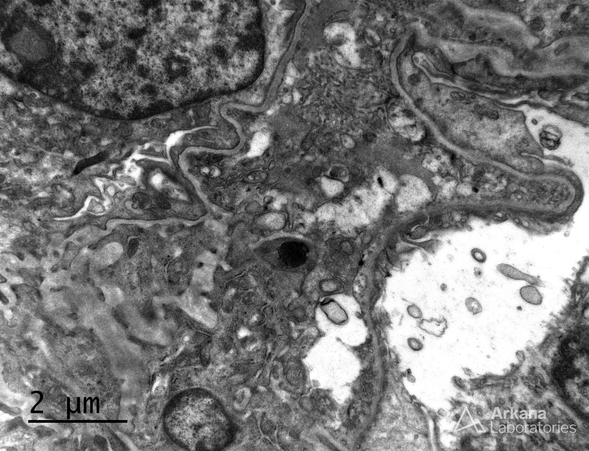
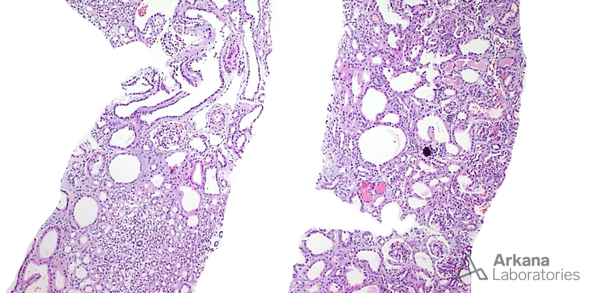
The morphological and electron microscopic features in this biopsy are suggestive of congenital nephrotic syndrome of the Finnish type (CNSF). CNSF is an autosomal recessive disease due to homozygous mutation of the NPHS1 gene on 19q31.1, which encodes for the protein nephrin that forms the ‘zipper’ structure of the slit diaphragm. Although it is found worldwide, Finlad has the highest incidence. CNSF accounts for approximately 60% of nephrotic syndromes in the first 3 months of life.
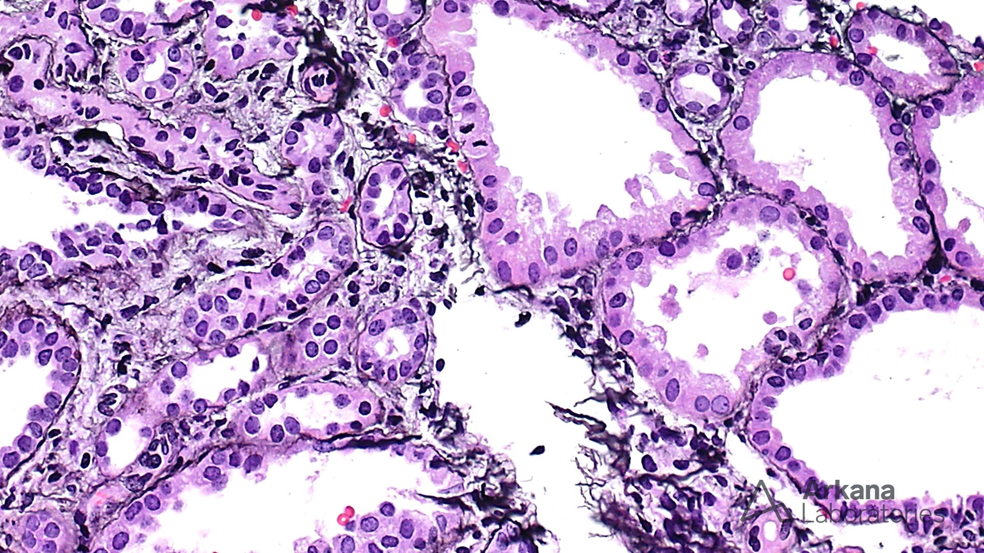
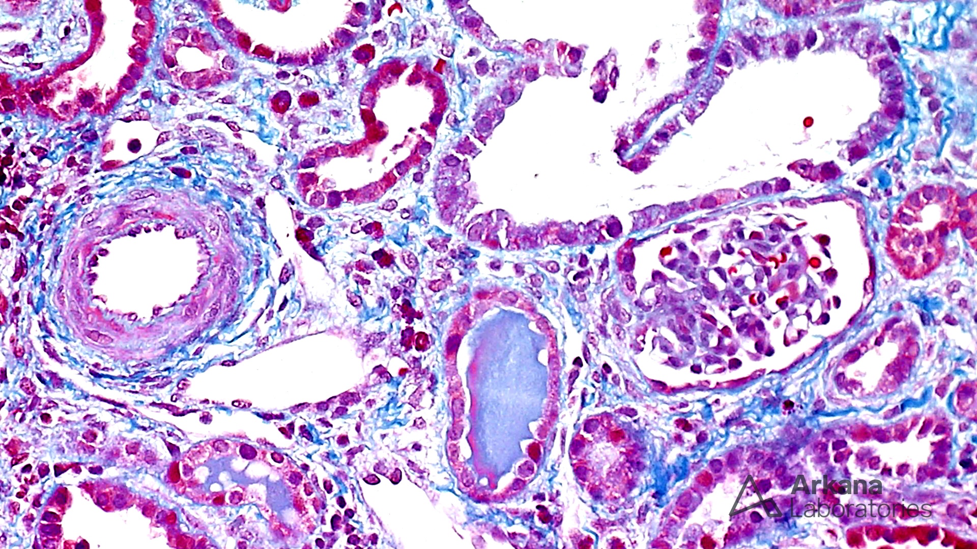
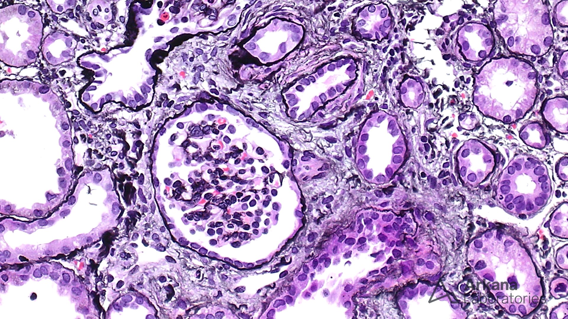
Mesangial sclerosis in CNSF is often less prominent than as seen in DMS. However, the low power impression of with many dilated tubules and thick walled blood vessels is a feature that is more commonly seen in CNSF. Denys Drash syndrome is a possibility, however, there is no evidence of pseudo-hermaphrodytism and no WT1 mutation. Moreover, in contrast to Denys Drash and Alport syndrome, no abnormalities in the thickness or contour of the basement membrane are seen in CNSF by electron microscopy. CNSF shows lack of the slit diaphragm. This is often difficult to view in a case like this when the foot processes are severely effaced. On the other hand, in minimal change disease, occasional filtration slit diaphragms can often be found. The presence of extra-glomerular features are also useful to differentiate from minimal change disease.
Quick note: This post is to be used for informational purposes only and does not constitute medical or health advice. Each person should consult their own doctor with respect to matters referenced. Arkana Laboratories assumes no liability for actions taken in reliance upon the information contained herein.