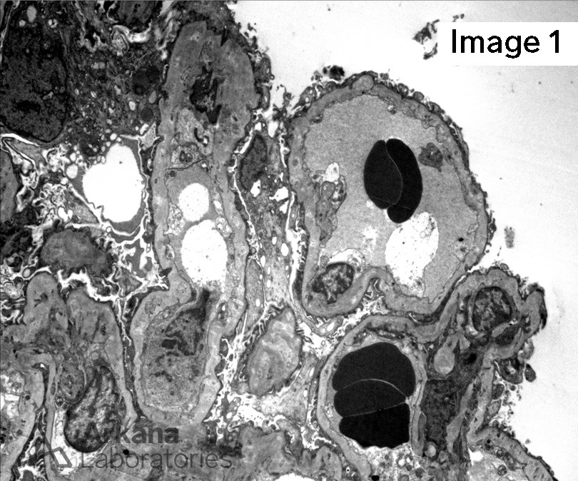
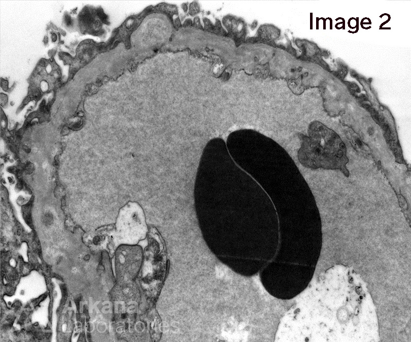
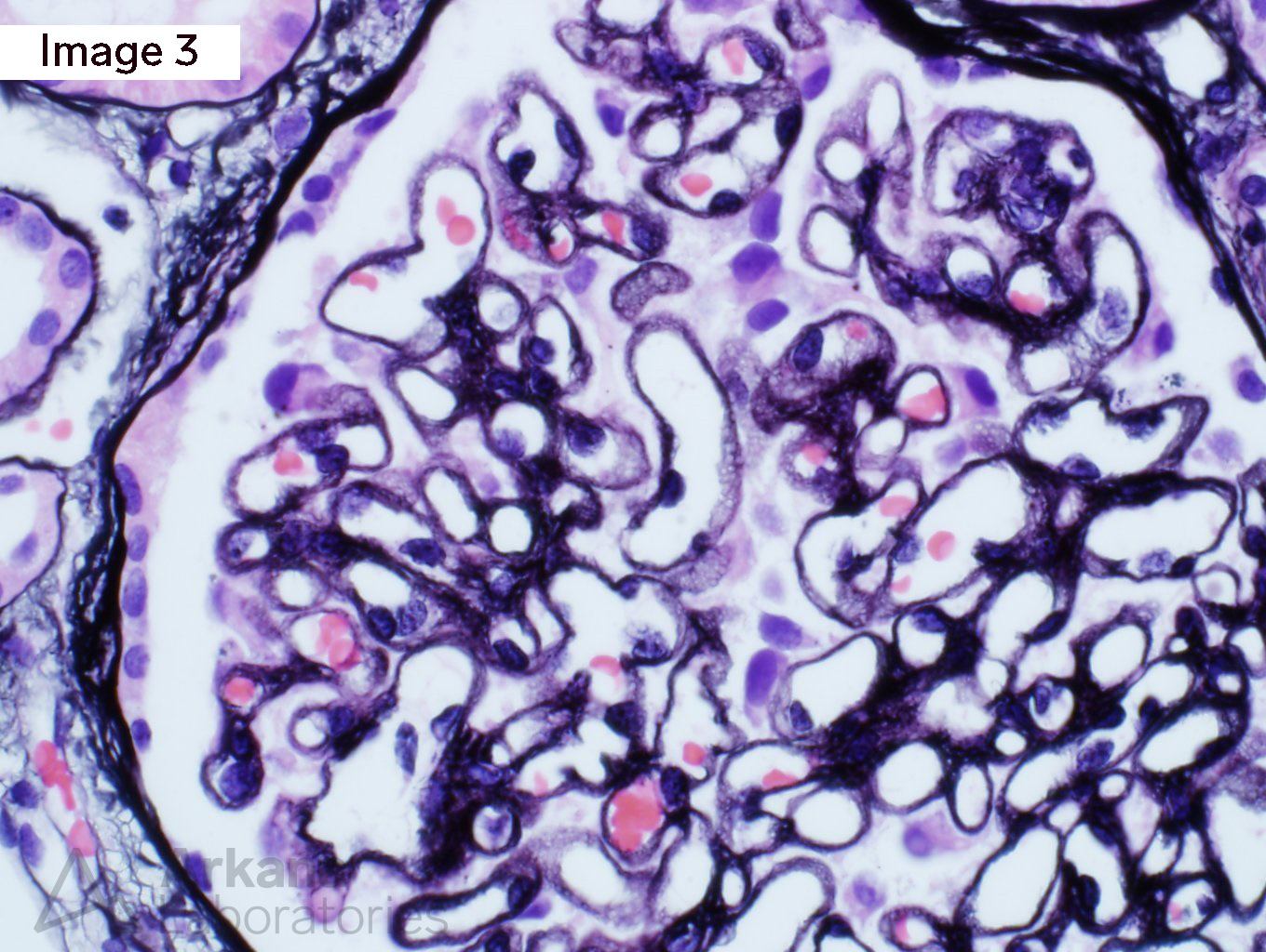
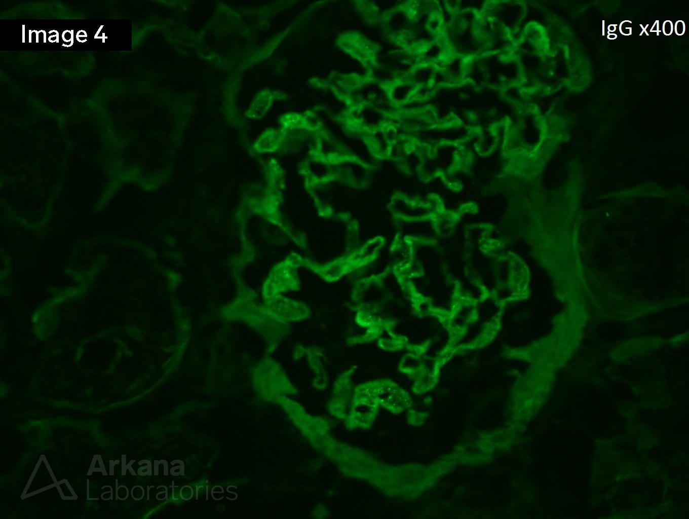
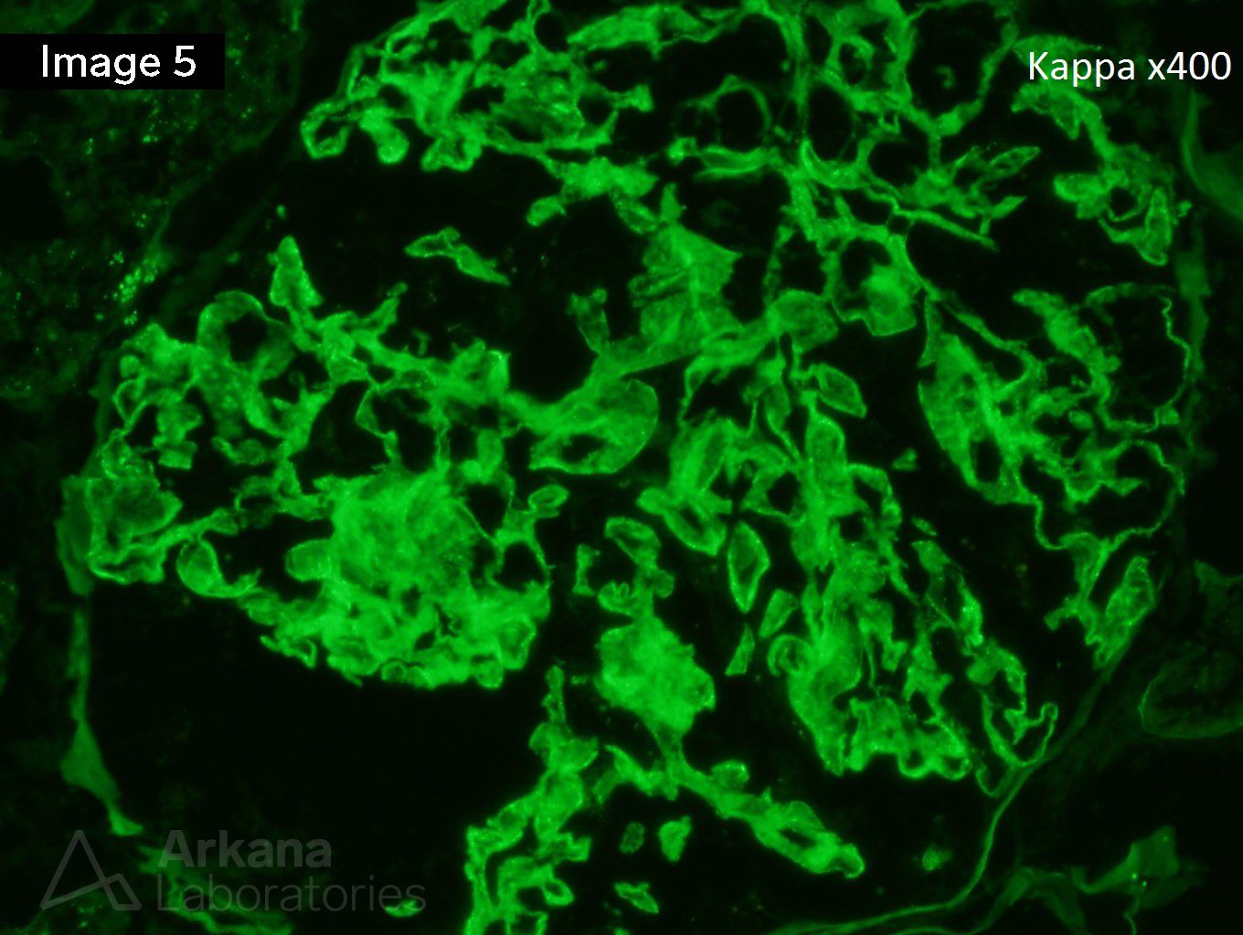
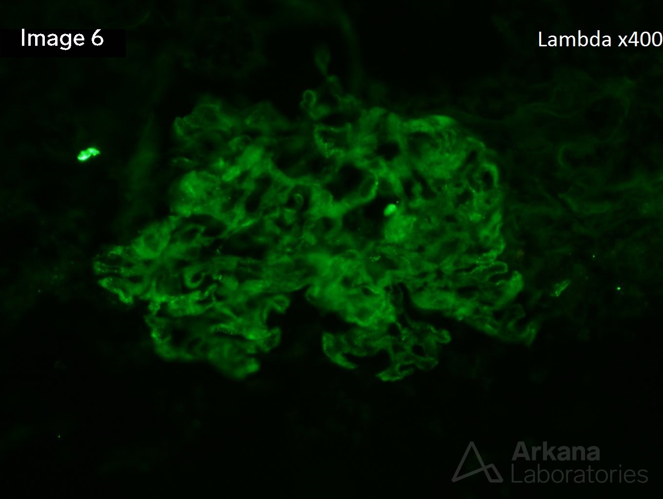
A 40-year-old white male presents with 3 grams/24 hr of proteinuria and swelling around his ankles. He reports that he has had foamy urine over the last 4 months, but was unable to go to a doctor till now because of lack of insurance. Image 1 and image 2 shows irregularly thickened glomerular basement membranes with lamellation. Image 3 shows numerous “holes” on silver stain. Images 4, 5, and 6 show weak staining for IgG, kappa, and lambda, respectively.
Membranous nephropathy in a resolving phase can be a tricky diagnosis since the immunofluorescence staining may be extremely weak or negative. The lamellation and reorganization of the glomerular basement membranes by electron microscopy can even give the appearance of Alport syndrome type changes. If you look very carefully, you can make out rare electron-dense subepithelial deposits and intramembranous electron lucency where deposits have already reabsorbed. This electron microscopic appearance would be classified as Stage 4 membranous nephropathy by Ehrenreich and Churg.
Quick note: This post is to be used for informational purposes only and does not constitute medical or health advice. Each person should consult their own doctor with respect to matters referenced. Arkana Laboratories assumes no liability for actions taken in reliance upon the information contained herein.