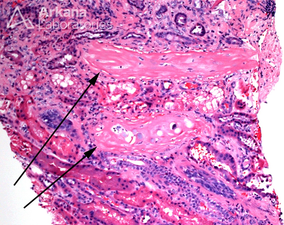
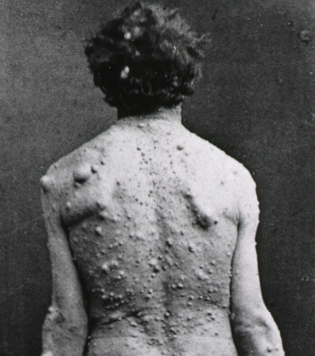
Today’s teaching point is a true “zebra” in renal pathology. The arrows in the light microscopic image identify what have been called renal sclerosing peritubular nodulerenal sclerosing peritubular nodules, which are foci of peritubular spindle cells with myofibroblastic differentiation and variable amounts of collagen formation. They are found in patients with neurofibromatosis type 2 (NF2). The black and white clinical photograph, in contrast, shows a patient with neurofibromatosis type 1 (NF1), in which renal sclerosing peritubular nodules have not been described. The etiology, classification, and clinical significance of these nodules remain unclear.
Gökden N et al. Renal sclerosing peritubular nodules in a patient with neurofibromatosis type 2: a case report with immunohistochemical and electron microscopic studies. Hum Pathol. 2009. PMID: 19540552.
Quick note: This post is to be used for informational purposes only and does not constitute medical or health advice. Each person should consult their own doctor with respect to matters referenced. Arkana Laboratories assumes no liability for actions taken in reliance upon the information contained herein.