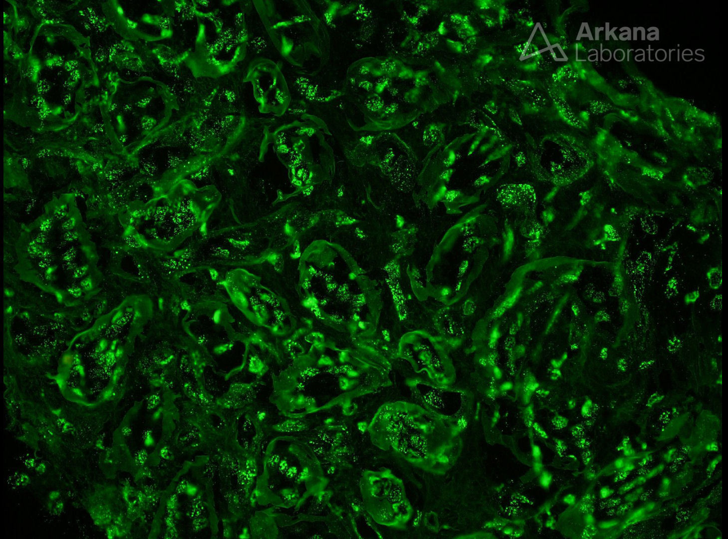
This immunofluorescence image (anti-IgG) shows prominent tissue ANA in a patient with lupus nephritis. Normally, the nuclei in renal tissues show no fluorescence signal for antibodies directed against immunoglobulin or complement components, so finding such staining is a helpful clue to the presence of underlying autoimmune disease (e.g. systemic lupus erythematosus). Staining includes homogeneous, speckled (shown in this case), or rim patterns, similar to that seen with serologic testing. For unclear reasons, however, tissue ANA is not always observed in patients with lupus — even in patients with positive ANA by serologic testing. For this reason, many consider this finding to represent a processing artifact instead of in vivo nuclear binding.
Quick note: This post is to be used for informational purposes only and does not constitute medical or health advice. Each person should consult their own doctor with respect to matters referenced. Arkana Laboratories assumes no liability for actions taken in reliance upon the information contained herein.