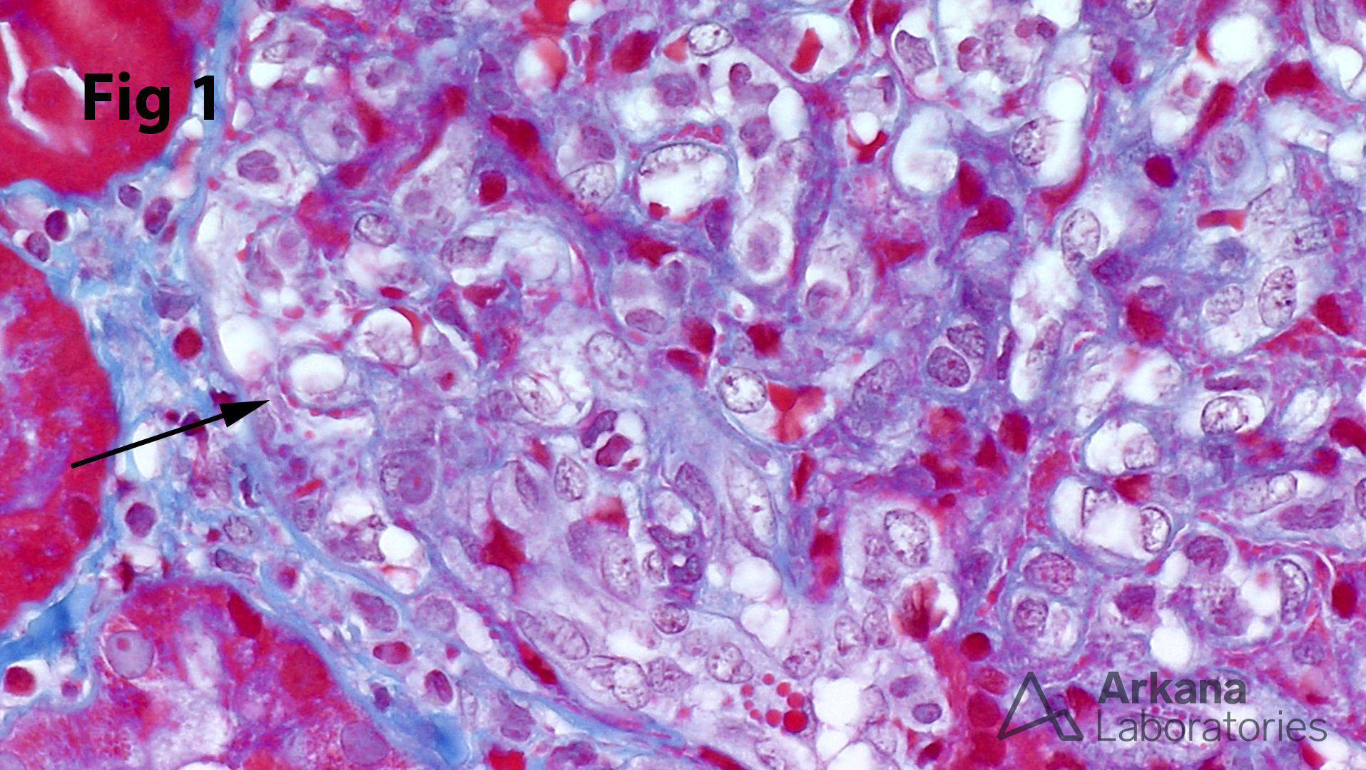
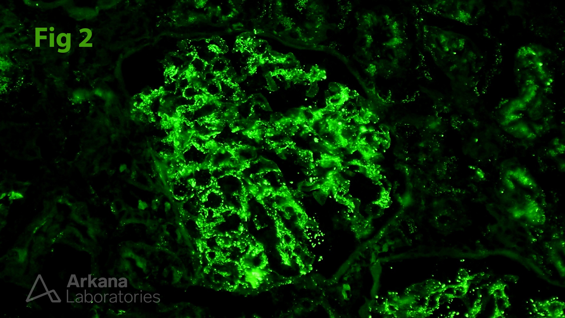
While the trichrome stain is best known for assessing the extent of interstitial fibrosis, it is a versatile stain with many applications. In this high-magnification image of a proliferative glomerulonephritis, the arrow points to red-pink (i.e. “fuchsinophilic”) subepithelial deposits along the blue-stained glomerular capillary wall. The corresponding immunofluorescence image from the same case shows extensive capillary wall IgG deposits (Fig. 2). If a particular biopsy lacks glomeruli or tissue for immunofluorescence studies, the trichrome stain can provide a useful clue that an immune complex-mediated disease process is present. In addition, the trichrome stain can be helpful in the identification of fibrin, both within glomeruli and in extraglomerular vessels. It even has characteristic staining patterns in various other glomerular deposits such as amyloid.
Quick note: This post is to be used for informational purposes only and does not constitute medical or health advice. Each person should consult their own doctor with respect to matters referenced. Arkana Laboratories assumes no liability for actions taken in reliance upon the information contained herein.