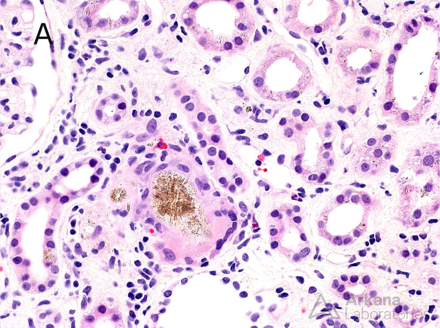
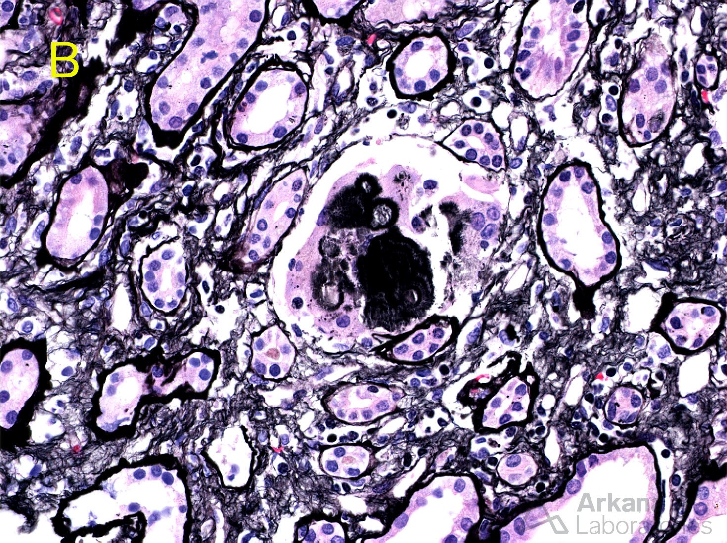
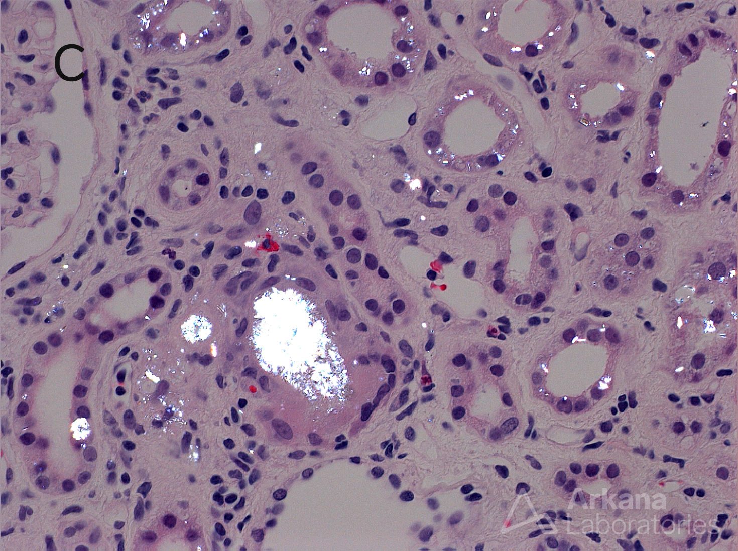
The renal biopsy shown here has crystals present within the tubular lumens and cytoplasm with a brownish appearance by H&E (A) that are strongly silver positive on the Jones methenamine silver stain (B). The crystals are birefringent when viewed under polarized light (C). These findings are characteristic of 2,8 DHA crystal deposition in the kidney resulting from a deficiency of adenine phosphoribosyltransferase. 2,8 dihydroxyadeninuria is an autosomal recessive disease resulting from pathogenic variants in the APRT gene. It is an important disease to recognize as treatment with a low purine diet and allopurinol therapy blocks formation of 2,8 DHA and can improve renal function. The differential diagnosis includes other crystalline nephropathies. One that is less well known but has a similar histopathology is triamterene crystalline nephropathy, which can be distinguished from 2,8 DHA by the presence of “Maltese crosses” under polarized light.
Quick note: This post is to be used for informational purposes only and does not constitute medical or health advice. Each person should consult their own doctor with respect to matters referenced. Arkana Laboratories assumes no liability for actions taken in reliance upon the information contained herein.