Clinical History:
- The patient is a 72-year-old man. Clinical diagnosis of neuropathy.
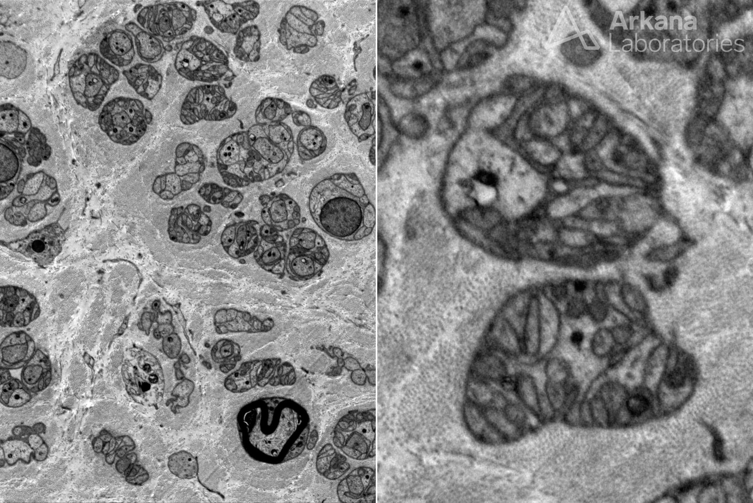
What cannot be seen on this electron micrograph of muscle?
A. Unmyelinated fiber loss
B. “Naked” axons
C. Large myelinated fiber loss
D. Small myelinated fiber loss
Answer:
“Naked axons”
This is a case of near total axonopathic fiber loss. “Naked axons” are generally large often dilated axons without Schwann cells investing them are only seen in active demyelinating disorders; no thinly myelinated axons, inflammation of macrophages extending into myelin layers are seen to corroborate a demyelinating syndrome.
Many Schwann cell subunits (cross sections of Schwann cell processes ”profiles” within a basement membrane) are present without obvious axons. Within these subunits, many Schwann cell profiles are seen which is a feature of small unmyelinated axon loss.
Reference(s) / Additional Reading:
- EM of small fiber changes
- Ochoa J. Muscle and Nerve 1978
- Small fiber loss
- Biopsy Diagnosis of Peripheral Neuropathy. G Midroni and JM Bilbao, editors. Butterworth-Heinemann Boston, 1995. 477 pp.
- Tavee J, Zhou L. Small fiber neuropathy. Cleve Clin J Med. 2009 May;76(5):297-305.
Quick note: This post is to be used for informational purposes only and does not constitute medical or health advice. Each person should consult their own doctor with respect to matters referenced. Arkana Laboratories assumes no liability for actions taken in reliance upon the information contained herein.