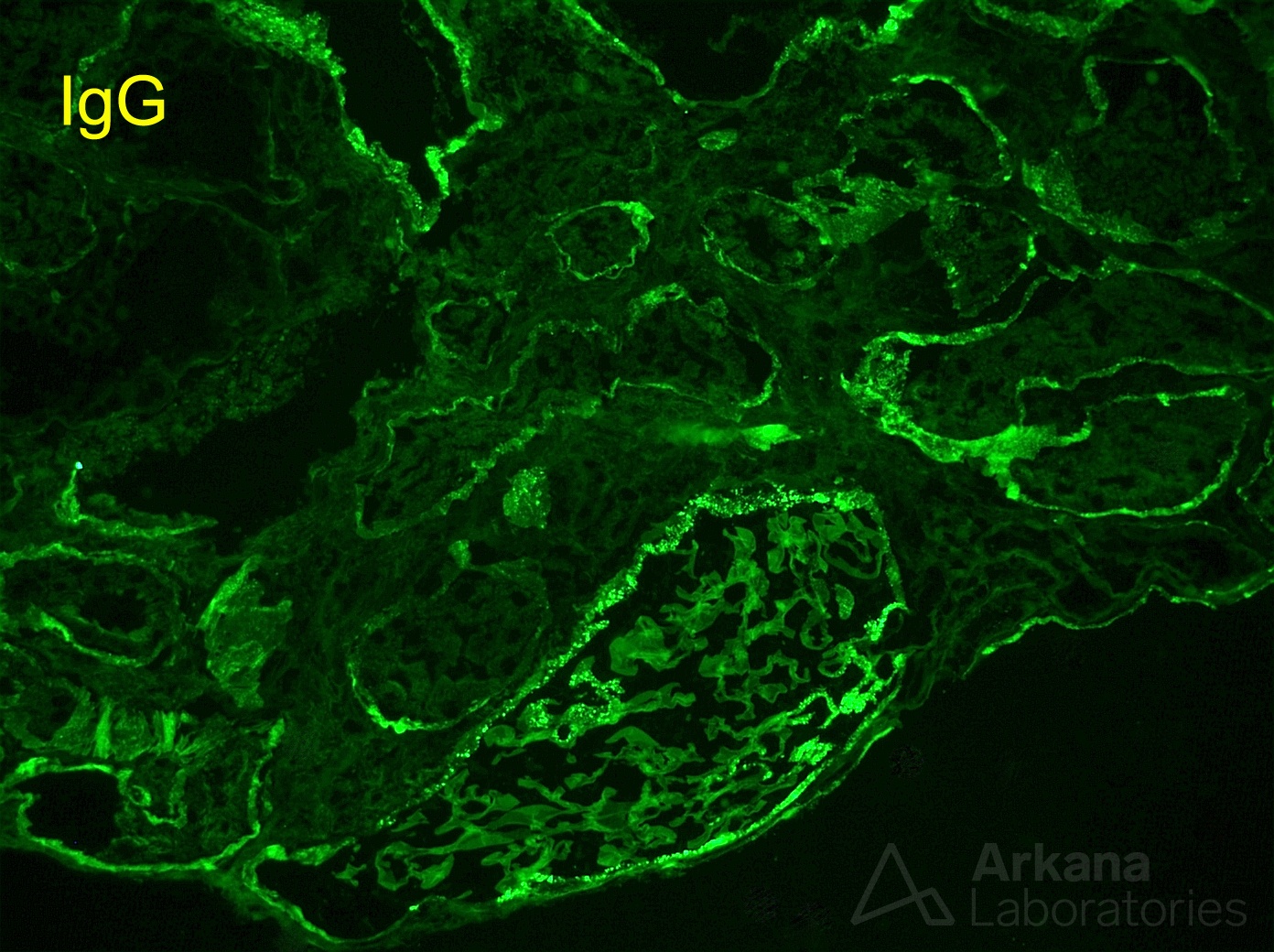
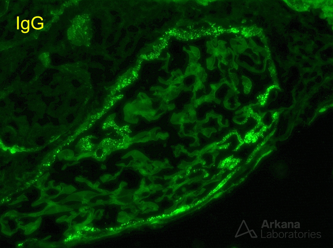
These photomicrographs show the classic immunofluorescence staining pattern for anti-LRP2 nephropathy, a disease that leads to severe renal tubular injury and is associated with circulating autoantibodies to the tubular brush border protein LRP2/megalin. The images show granular IgG staining along proximal tubular basement membrane, Bowman’s capsule, and segmental glomerular basement membrane staining. The presence of IgG-containing TBM immune deposits, particularly in elderly patients, should alert the clinician and pathologist to the possibility of this disorder. Similar tubular deposits can be seen in the setting of lupus nephritis and IgG4-related disease. Tubular deposits in the setting of lupus generally correlate with the presence of proliferative glomerular lesions, which was not present in this case by light microscopy. IgG4-related tubulointerstitial nephritis is the entity with the most morphologic overlap, as both it and anti-LRP2 nephropathy can exhibit interstitial inflammation and glomerular deposits in addition to the tubular deposits. However, most cases of IgG4-related disease show intense interstitial inflammation while inflammation in anti-LRP2 nephropathy was absent to mild in most cases.
Reference: Larsen CP, Trivin-Avillach C, Coles P, Collins AB, et al. LDL Receptor-Related Protein 2 (Megalin) as a Target Antigen in Human Kidney Anti-Brush Border Antibody Disease. J Am Soc Nephrol. 2017 Oct 26. [Epub ahead of print]
Quick note: This post is to be used for informational purposes only and does not constitute medical or health advice. Each person should consult their own doctor with respect to matters referenced. Arkana Laboratories assumes no liability for actions taken in reliance upon the information contained herein.