
Clinical presentation:
A 13-year-old male with no significant medical history presents with sudden onset nephrotic syndrome. His blood pressure is 160/90. He has 7.0 grams proteinuria with hypoalbuminemia, 1.0 mg/dL. He also has elevated triglycerides.
What is the differential for nephrotic syndrome in children?
The major causes of nephrotic syndrome in children are minimal change disease, focal segmental glomerulosclerosis (FSGS), and membranous nephropathy.
Now let’s look at the biopsy. Figure 1 shows the main histopathologic finding (Jones silver stain).
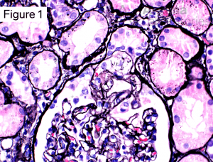
How would you describe this lesion?
There is segmental sclerosis of the glomerular tuft, and the area of sclerosis involves the urinary pole at the origin of the proximal tubule.
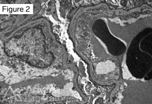
Our final clues are global podocyte foot effacement by electron microscopy (Figure 2) and negative immunofluorescence within the glomeruli.
What is the diagnosis based on the biopsy findings?
The pattern of glomerular injury seen in the biopsy, taken together with the clinical presentation makes the diagnosis Focal Segmental Glomerulosclerosis (FSGS) Tip Variant. Focal segmental glomerulosclerosis is a pattern of injury with segmental scar involving some but not all glomeruli. The patient’s presentation with nephrotic syndrome and extensive podocyte foot process effacement support a primary podocytopathy (i.e. FSGS due to podocyte injury). There is a morphologic spectrum of FSGS. The Columbia classification groups FSGS into five histologic variants. In the tip variant, a segmental lesion located at the outer 25% of the glomerular tuft with adhesion to Bowman’s capsule adjacent to the proximal tubule or a predominance of podocytes at the proximal tubular lumen is sufficient for diagnosis. There is usually endocapillary hypercellularity. Sclerosis and foam cells may also be present. In particular, the diagnosis of the tip variant requires exclusion of a collapsing lesion.
References
Jennette JC, Olson JL, Silva FG, D’Agati V, Heptinstall RH *1920 *, eds. Heptinstall’s Pathology of the Kidney: Includes Interactive EBook with Complete Content. Seventh edition. Wolters Kluwer; 2015.
D’Agati VD, Fogo AB, Bruijn JA, Jennette JC. Pathologic classification of focal segmental glomerulosclerosis: a working proposal. American Journal of Kidney Diseases. 2004;43(2):368-382.
Quick note: This post is to be used for informational purposes only and does not constitute medical or health advice. Each person should consult their own doctor with respect to matters referenced. Arkana Laboratories assumes no liability for actions taken in reliance upon the information contained herein.