
The patient is a 75-year-old female with a history of hypertension who presents with acute kidney injury. Her serum creatinine is 12 mg/dL and her urine protein creatinine ratio is 17.
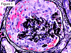
The main finding on kidney biopsy is shown below (Jones silver stain).
What is this lesion?
This is a glomerular crescent. A crescent is an extra capillary proliferation usually associated with fibrin and fibrous matrix. The amount of cells/fibrin in relation to the fibrous matrix determines the type of crescent. A cellular crescent has more than 75% cells/fibrin. A fibrous crescent has more than 75% fibrous matrix, and a fibrocellular crescent has 25-75% cells/fibrin and the remainder fibrous matrix. In this case, all glomeruli showed involvement by cellular crescents. Note the area of fibrinoid necrosis.
What is the differential for crescents?
The finding of glomerular crescents has three main etiologies
1. Anti-Glomerular Basement Membrane (Anti-GBM) Glomerulonephritis
2. Pauci-immune glomerulonephritis, and
3. Immune complex glomerulonephritis (such as Lupus nephritis, IgA nephritis).
Additional clues
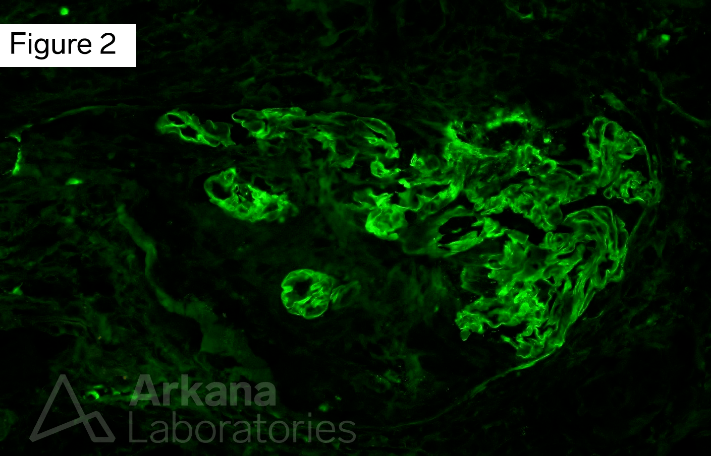
The final clues are seen on immunofluorescence. There is global linear capillary wall staining for IgG (Figure 2). Similar staining is seen for Kappa and Lambda. Additionally, the fibrinogen stain highlights fibrin in areas of fibrinoid necrosis (Figure 3). All other stains are negative.

What is the diagnosis based on the biopsy findings?
The diagnosis is anti-glomerular basement membrane glomerulonephritis. Glomerular crescents are a pathologic change that indicate severe glomerular injury. The finding of crescents that diffusely involve glomeruli should raise suspicion for anti-GBM glomerulonephritis. Staining for immunoglobulins can help further determine the cause of the glomerular injury. Anti-GBM glomerulonephritis shows linear GBM staining for IgG. Pauci-immune glomerulonephritis has minimal to no staining for IgG and immune complex glomerulonephritis typically has a granular staining pattern for IgG.
References
Jennette JC, Olson JL, Silva FG, D’Agati V, Heptinstall RH *1920 *, eds. Heptinstall’s Pathology of the Kidney: Includes Interactive EBook with Complete Content. Seventh edition. Wolters Kluwer; 2015.
Quick note: This post is to be used for informational purposes only and does not constitute medical or health advice. Each person should consult their own doctor with respect to matters referenced. Arkana Laboratories assumes no liability for actions taken in reliance upon the information contained herein.