This 9-year-old female was recently evaluated for lymphoid malignancy due to weight loss and intermittent fevers. Following a negative workup for malignancy, further routine studies showed mild hematuria and proteinuria and she was referred to a pediatric nephrologist. The serologic evaluation showed a positive ANA and positive double-stranded DNA, complement C3 borderline low and C4 normal. Urinalysis showed 3-5 RBCs/HPF but no casts with 2+ blood and 1+ protein. Mesangial lupus was suspected but a biopsy was done to rule out activity.
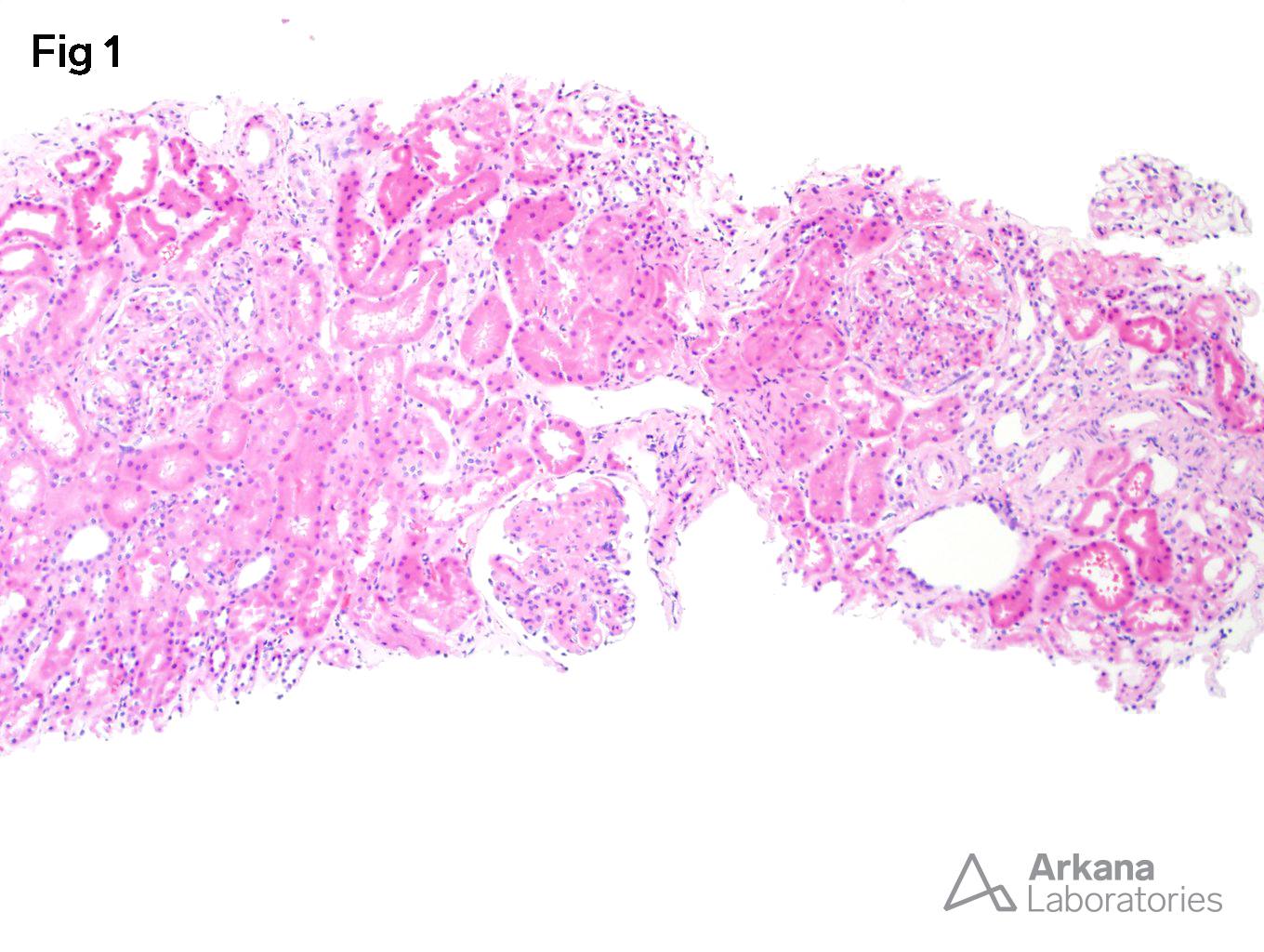
Figure 1 – No fibrosis with diffuse mesangial hypercellularity and focal proliferation but no crescents and no hyaline thrombi (H&E 100x).
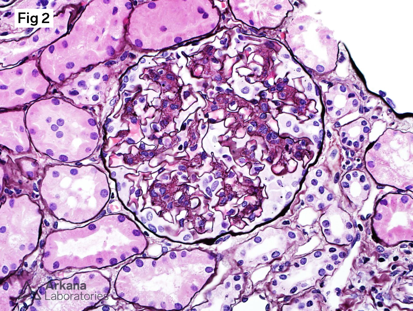
Figure 2 – Mesangial matrix expansion and mesangial hypercellularity (Jones silver 400x).
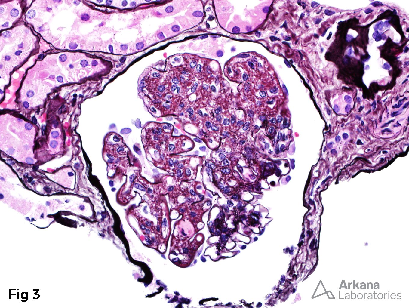
Figure 3 – Endocapillary proliferation in more than three-quarters of the glomerulus. Note the open loops at around 5:00 o’clock (Jones silver 400x).
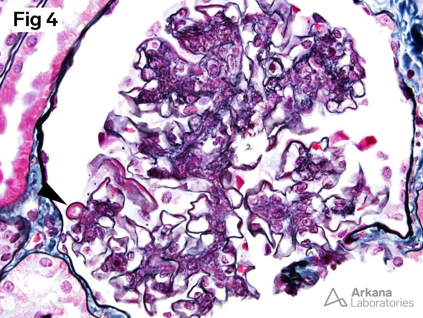
Figure 4 -Wire loop (arrowhead) in a glomerulus with only mesangial hypercellularity (Silver Methenamine Masson Trichrome or SMMT 400x).
Diagnosis: Focal Lupus Nephritis, Class III (A) with mild activity and no chronicity
Quick note: This post is to be used for informational purposes only and does not constitute medical or health advice. Each person should consult their own doctor with respect to matters referenced. Arkana Laboratories assumes no liability for actions taken in reliance upon the information contained herein.