What is your diagnosis?
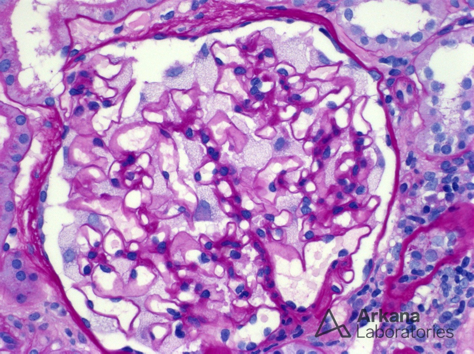
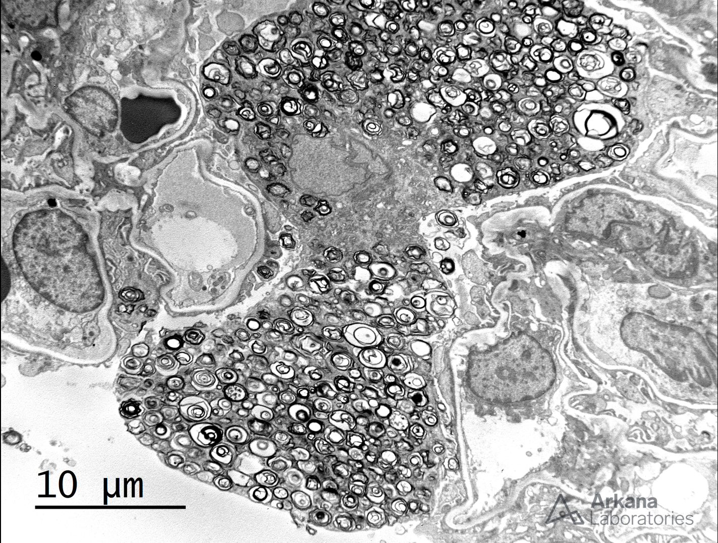
This biopsy shows a glomerulus with podocytes displaying extensively vacuolated cytoplasm. While this is a non-specific finding, it is very uncommon and most frequently seen in the setting of Fabry disease. Fabry disease is an X-linked disorder caused by a deficiency of the lysosomal enzyme α-galactosidase A, which catalyzes the breakdown of glycosphingolipids. Deficiency of this enzyme results in lysosomal accumulation of globotriaosylceramide in many organs and within the kidney, globotriaosylceramide may accumulate within endothelial cells, podocytes, distal tubular cells and collecting duct epithelium. Identification of myeloid bodies within podocytes by electron microscopy (see image below) is highly suggestive, but not diagnostic of the disease, given that similar inclusions may be seen in patients treated with amiodarone, chloroquine, and hydroxychloroquine. Correlation with serum testing for α-galactosidase A levels is useful for definitive diagnosis.
Quick note: This post is to be used for informational purposes only and does not constitute medical or health advice. Each person should consult their own doctor with respect to matters referenced. Arkana Laboratories assumes no liability for actions taken in reliance upon the information contained herein.