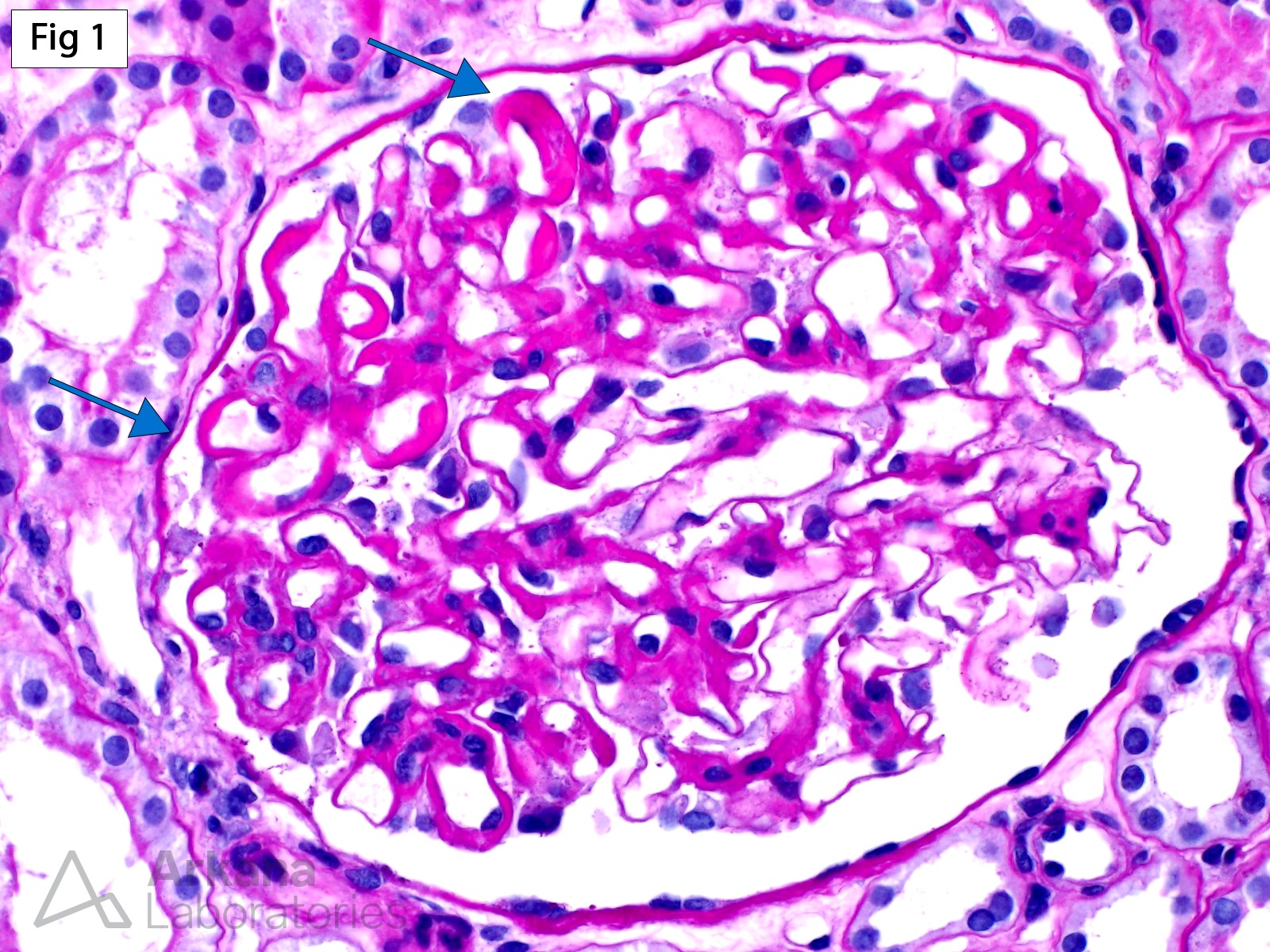
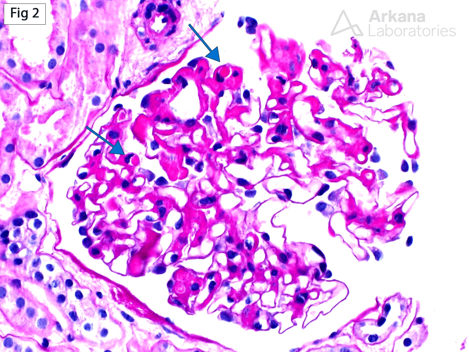
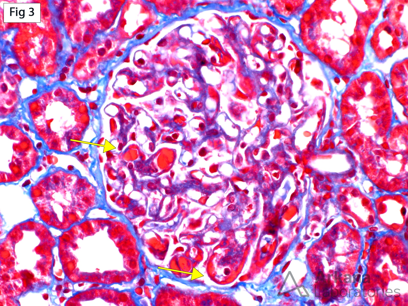
The ISN/RPS 2004 classification of lupus nephritis is entirely based on glomerular changes. Depending on the presence or absence of active and/or chronic glomerular lesions, cases are allocated into one (and occasionally two) of six possible classes. The most commonly encountered active glomerular lesions in clinical practice are in the form of endocapillary or extracapillary proliferation, fibrinoid necrosis or cellular crescents. However, deposition of large subendothelial immune deposits, even in the absence additional proliferative/necrotizing lesions is sufficient to classify a case as class III or IV, depending on the number of involved glomeruli (Fig 1-3). These immune deposits, termed “wire loops”, appear as glassy eosinophilic, strongly PAS-positive, fuchsinophilic and non-argyrophilic material, which involves the circumference of a capillary loop (Fig 1 & Fig 3). When these same deposits extend into the lumen of a loop and are sectioned en face, they appear as endocapillary thrombi and are thus called “hyaline thrombi” (Fig 2). The nature of these deposits may be further confirmed by immunofluorescence and/or electron microscopy.
Quick note: This post is to be used for informational purposes only and does not constitute medical or health advice. Each person should consult their own doctor with respect to matters referenced. Arkana Laboratories assumes no liability for actions taken in reliance upon the information contained herein.