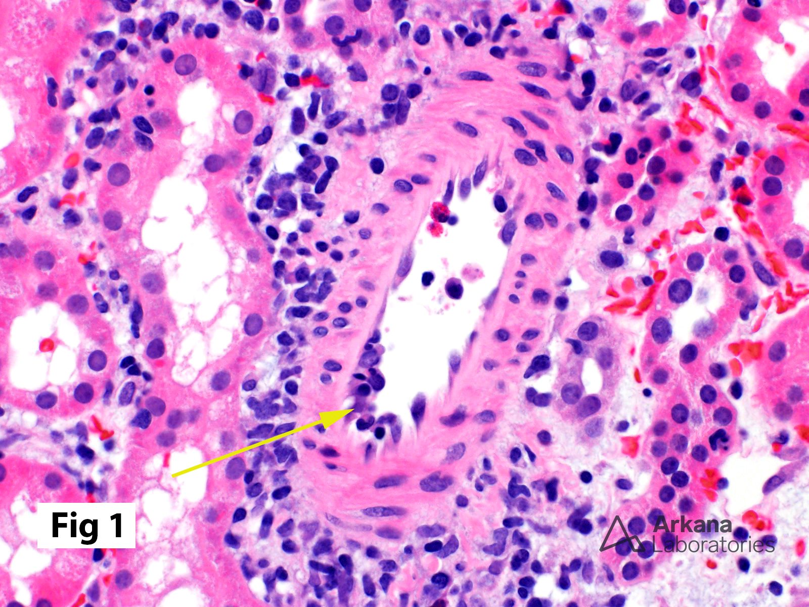
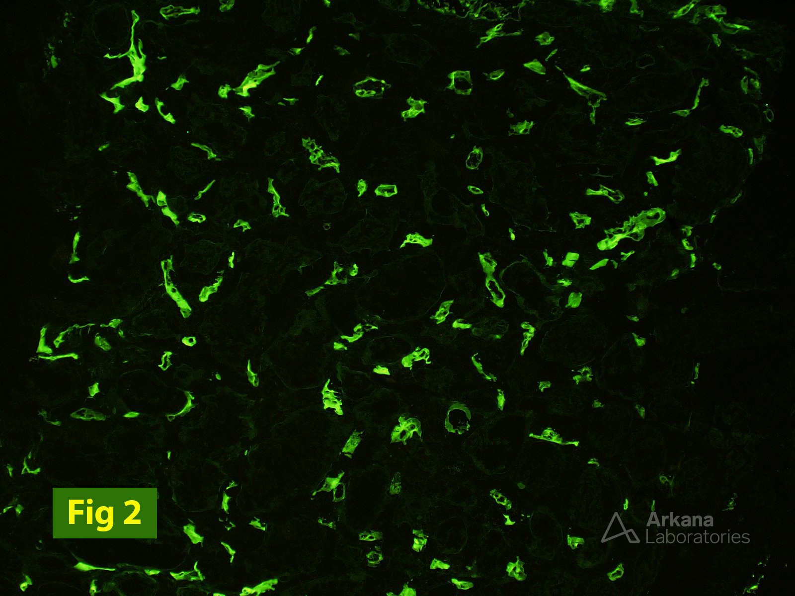
The image in Figure 1 shows mild intimal arteritis in an allograft biopsy from a patient who had undergone a kidney transplant. Note that the mononuclear inflammatory cells are found beneath the endothelium rather than simply adherent to the endothelial surface, which may be seen with inflammatory cell margination. Remember, too, as reflected in the 2013 revised Banff working classification, that intimal arteritis may be seen in antibody-mediated rejection in addition to T-cell-mediated rejection. In the patient’s biopsy, the additional presence of linear C4d positivity in peritubular capillaries (Figure 2) and the absence of significant interstitial inflammation and tubulitis provided strong morphologic evidence supporting acute/active antibody-mediated rejection.
Reference:
Haas M, et al. Banff 2013 meeting report: inclusion of c4d-negative antibody-mediated rejection and antibody-associated arterial lesions. Am J Transplant. 2014 Feb;14(2):272-83. PubMed PMID: 24472190.
[https://www.ncbi.nlm.nih.gov/pubmed/24472190]
Quick note: This post is to be used for informational purposes only and does not constitute medical or health advice. Each person should consult their own doctor with respect to matters referenced. Arkana Laboratories assumes no liability for actions taken in reliance upon the information contained herein.