When giant cells surround tubules…
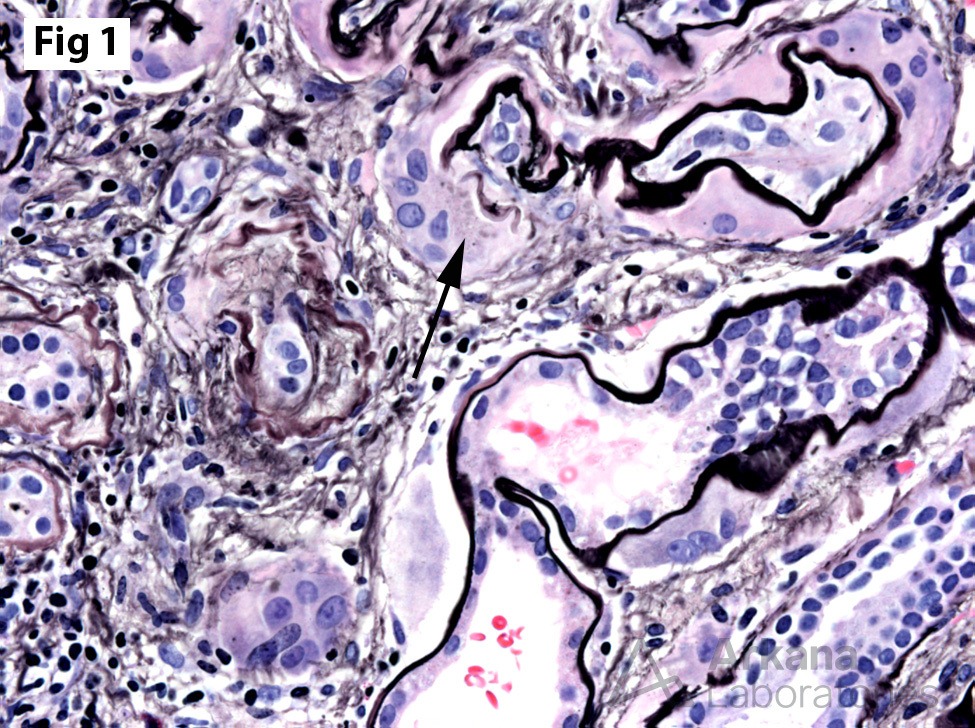
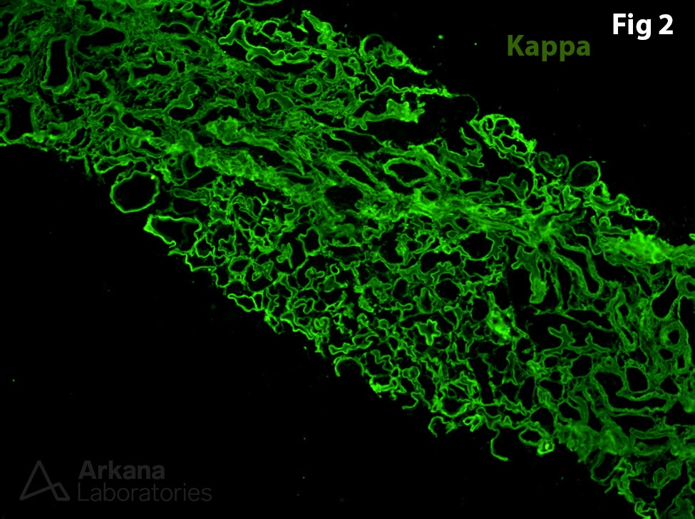
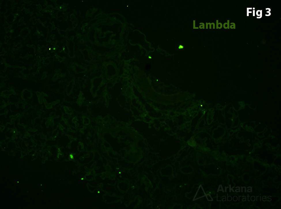
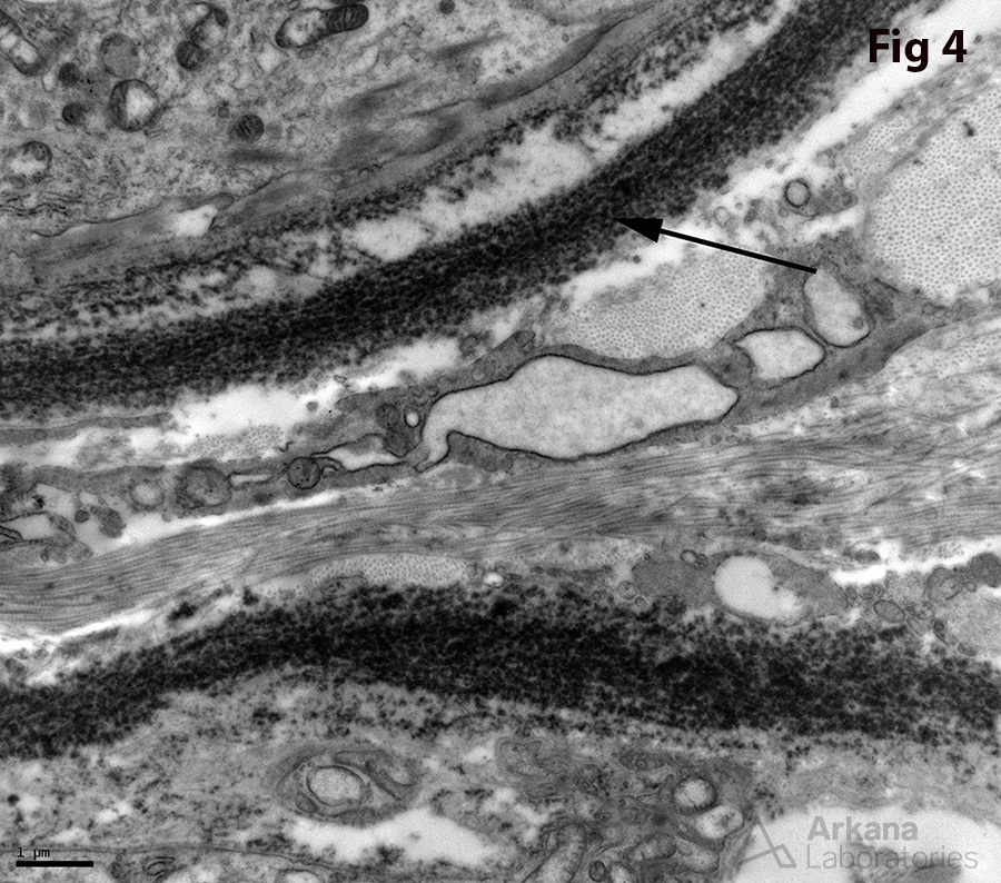
This renal biopsy was taken from a young woman with elevated serum creatinine (4.3 mg/dL), hematuria, and proteinuria. By light microscopy, there is a prominent giant cell reaction surrounding many of the tubules (Fig 1), the differential diagnosis of which includes so-called giant cell tubulitis (a form of tubulointerstitial nephritis associated with tubular basement membrane immune complex deposits), lupus tubulointerstitial nephritis, and monoclonal immunoglobulin deposition disease. By immunofluorescence, there is intense positivity of the tubular and glomerular basement membranes for kappa light chain (3+) with no corresponding staining for lambda light chain (Fig 2-3). The electron microscopic image shows fine granular deposits along the tubular basement membranes (Fig 4). These findings support the diagnosis of kappa light chain deposition disease with associated peritubular giant cells.
Yamashita F, et al. Light chain nephropathy with a remarkable accumulation of multinucleated giant cells in the kidney. Nihon Jinzo Gakkai Shi. 1994 Nov;36(11):1276-81. PMID: 7853760.
Quick note: This post is to be used for informational purposes only and does not constitute medical or health advice. Each person should consult their own doctor with respect to matters referenced. Arkana Laboratories assumes no liability for actions taken in reliance upon the information contained herein.