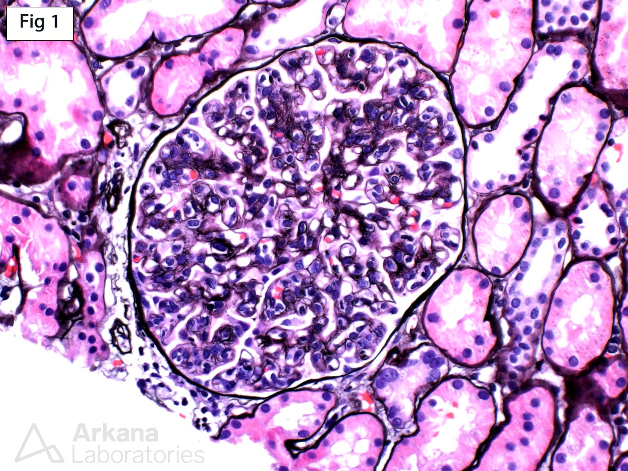
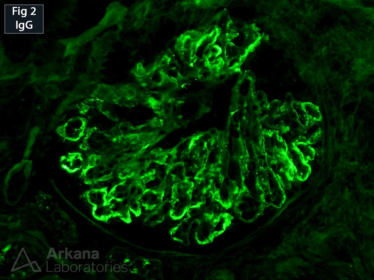
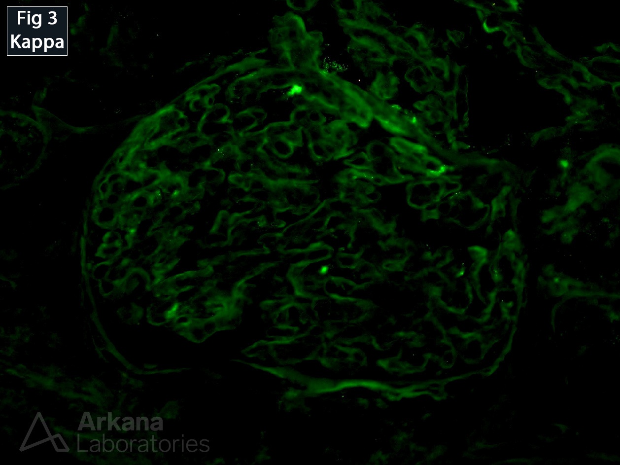
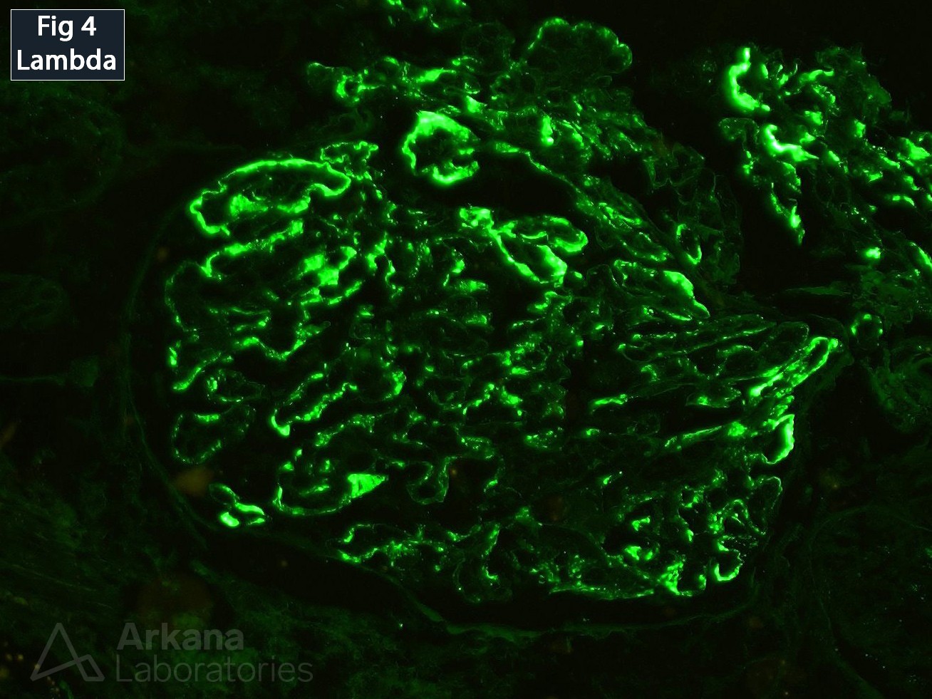
This biopsy was performed on a 58-year-old female who presented with mild proteinuria. The glomeruli show diffuse and global endocapillary hypercellularity with segmental double contour formation of the capillary loops on the silver stain (Fig 1). Immunofluorescence shows mesangial and segmental capillary wall deposits positive for IgG (3+, Fig 2), C3 (3+, not shown), C1q (1+, not shown) and lambda light chain (3+, Figure 4). Staining for kappa light chain is negative in glomeruli (Fig 3). Further immunofluorescence staining for IgG subclasses proves the deposits to be IgG3 restricted. Electron microscopy shows mesangial and segmental subendothelial immune-complex type deposits. The overall findings are consistent with the entity known as proliferative glomerulonephritis with monoclonal IgG deposits (see reference). While the deposits are most commonly IgG3 κ restricted (53.1%), cases with IgG3 λ deposits (12.5%) have also been reported.
Reference: Nasr S, Satoskar A, et al. Proliferative Glomerulonephritis with Monoclonal IgG Deposits. J Am Soc Nephrol. 20:2055-2064, 2009.
Quick note: This post is to be used for informational purposes only and does not constitute medical or health advice. Each person should consult their own doctor with respect to matters referenced. Arkana Laboratories assumes no liability for actions taken in reliance upon the information contained herein.