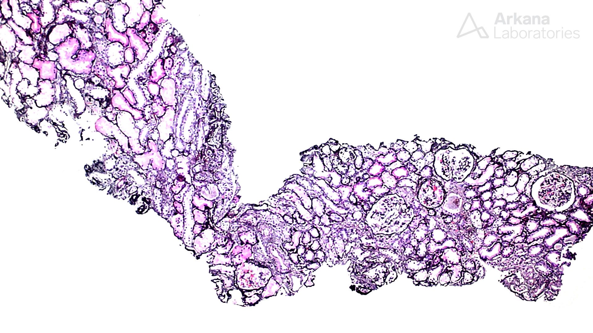
These images are from the kidney biopsy of a 70-year-old woman with a history of hypertension and valvular heart disease presenting with renal failure and serum creatinine of 4 mg/dl. The findings are suggestive of a mutation in which of the following genes:
A. Podocin
B. Glucocerebrosidase
C. a-galactosidase A
D. Nephrin
E. Phospholipase C

Answer: C
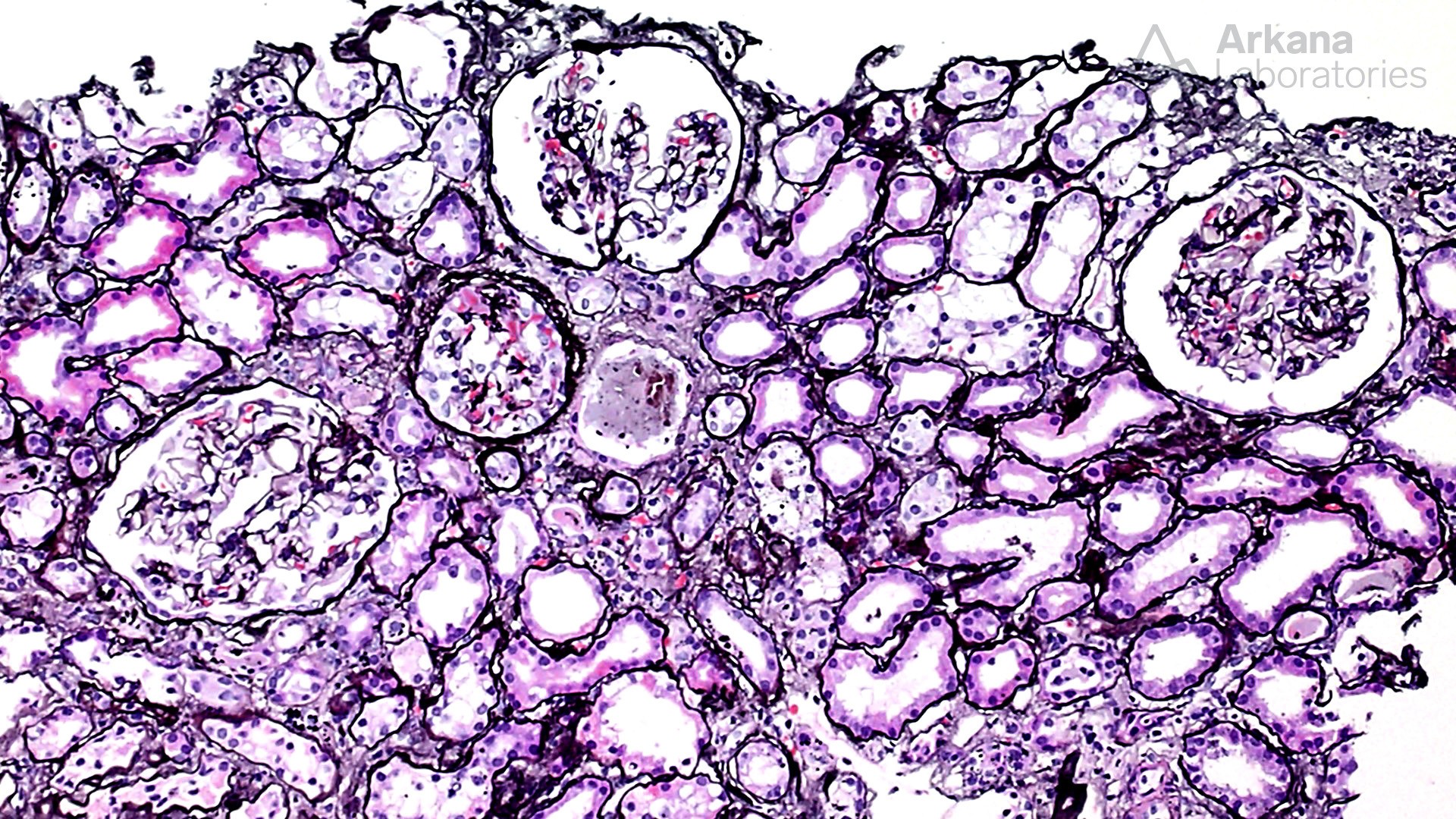
The images highlight podocytes loaded with numerous myelinosomes. There are only a few causes that can cause such accumulation, most notably Fabry disease. However, rare secondary causes to be considered include drugs such as chloroquine and chloroquine-like drugs (e.g., Plaquenil), amiodarone, aminoglycoside antibiotics, antidepressants and anti-cholesterol medications.
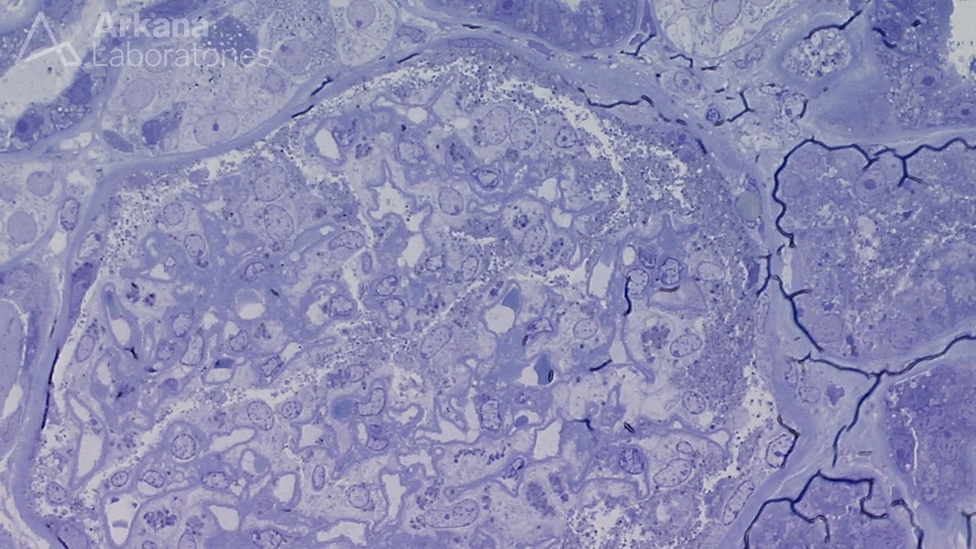
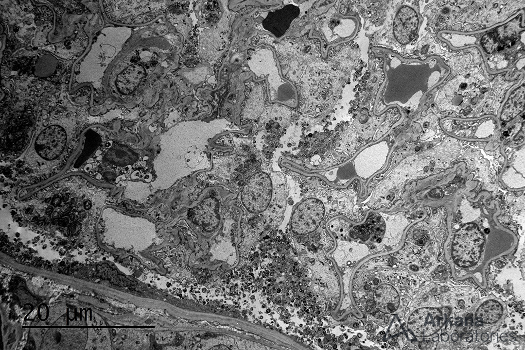
Fabry’s disease is an X-linked recessive, lysosomal storage disease caused by mutations in the gene a-galactosidase A (GLA) on the long arm of chromosome X (Xq22.1). The mutations lead to a deficiency of the enzyme a-galactosidase A and accumulation of globotriaosylceramide (GL3) in different cells including myocytes, podocytes, endothelial and distal tubules. The lipid-containing myelinosomes can be appreciated in the toluidine blue sections because osmium fixation preserves the lipid. By electron microscopy laminated, electron-dense deposits can be seen, which have been classically described as “zebra bodies”. Females are heterozygous carriers and many (30%) are asymptomatic.
Quick note: This post is to be used for informational purposes only and does not constitute medical or health advice. Each person should consult their own doctor with respect to matters referenced. Arkana Laboratories assumes no liability for actions taken in reliance upon the information contained herein.