This biopsy is taken from a 39-year-old woman who presents with abdominal pain, ascites, and lower extremity edema. Her serum creatinine is 3.9 mg/dL and her complete blood count shows leukocytosis (14,500). Initial serologic workup is negative.
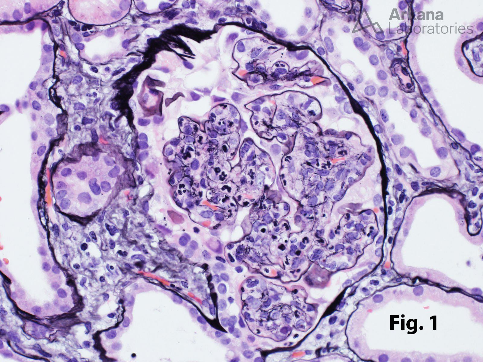
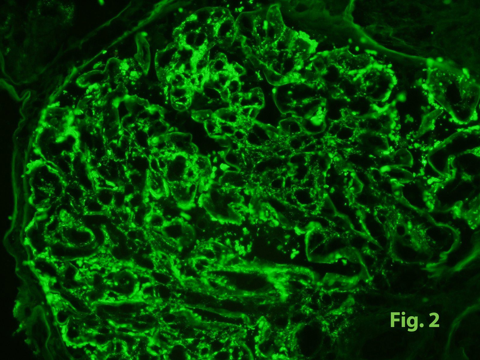
The biopsy shows a diffuse proliferative glomerulonephritis characterized by global endocapillary hypercellularity with prominent neutrophils, best visualized using methenamine silver staining (Fig. 1). No crescents, necrotizing lesions, or significant double contours are identified. By immunofluorescence, there is coarsely granular (3+) capillary wall and less prominent mesangial staining for IgG, C3, kappa, and lambda (Fig. 2). Electron microscopy shows global endocapillary proliferation and numerous large hump-like subepithelial electron dense deposits along with smaller subendothelial and mesangial deposits.
The pathologic findings are most consistent with infection-associated renal disease, so additional clinical workup for an underlying infectious etiology was initiated.
Quick note: This post is to be used for informational purposes only and does not constitute medical or health advice. Each person should consult their own doctor with respect to matters referenced. Arkana Laboratories assumes no liability for actions taken in reliance upon the information contained herein.