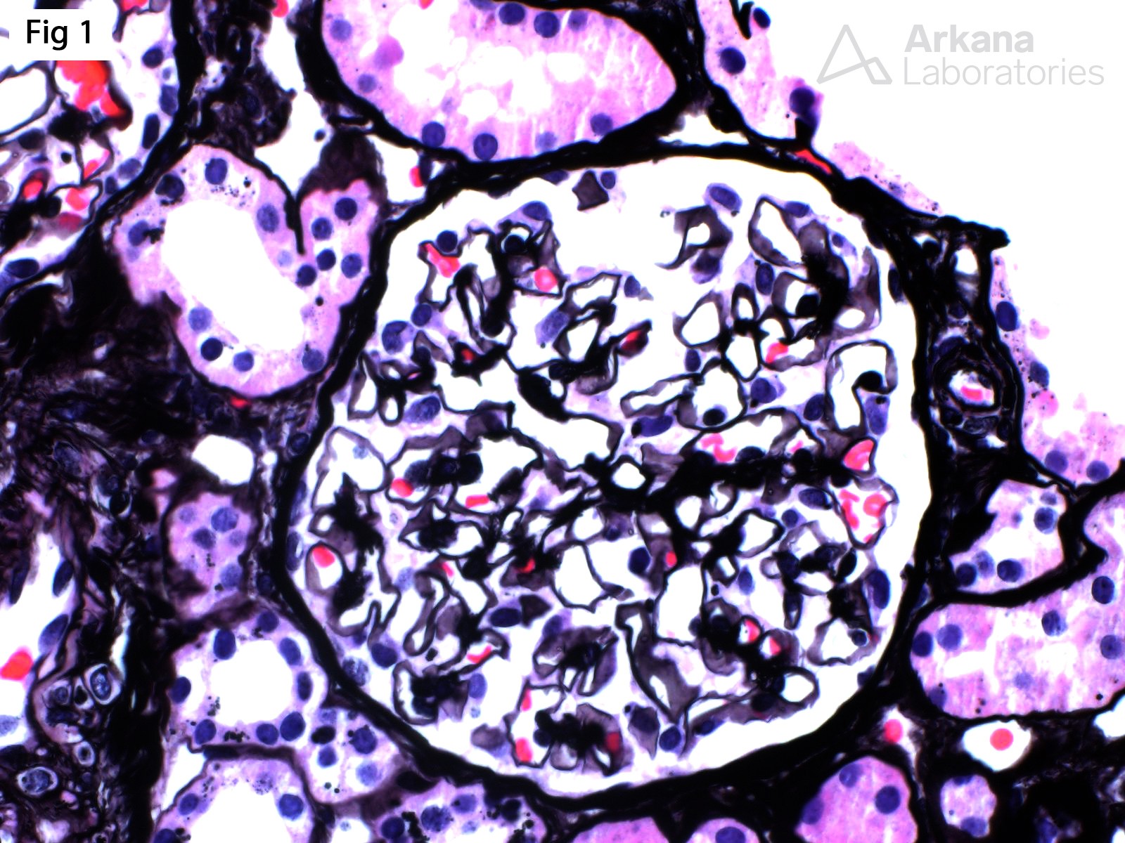
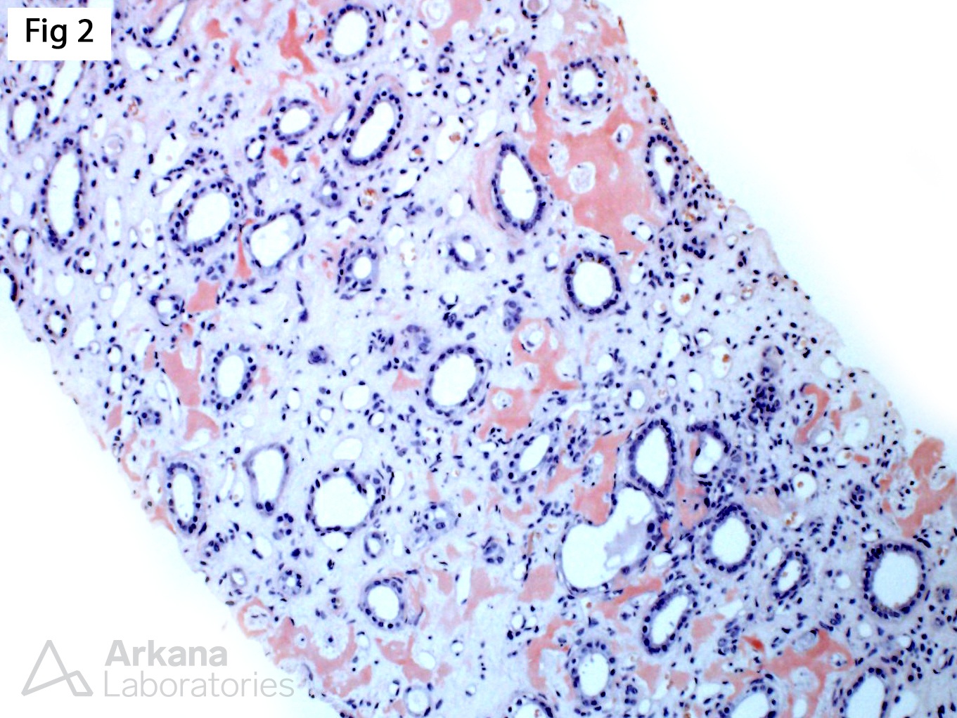
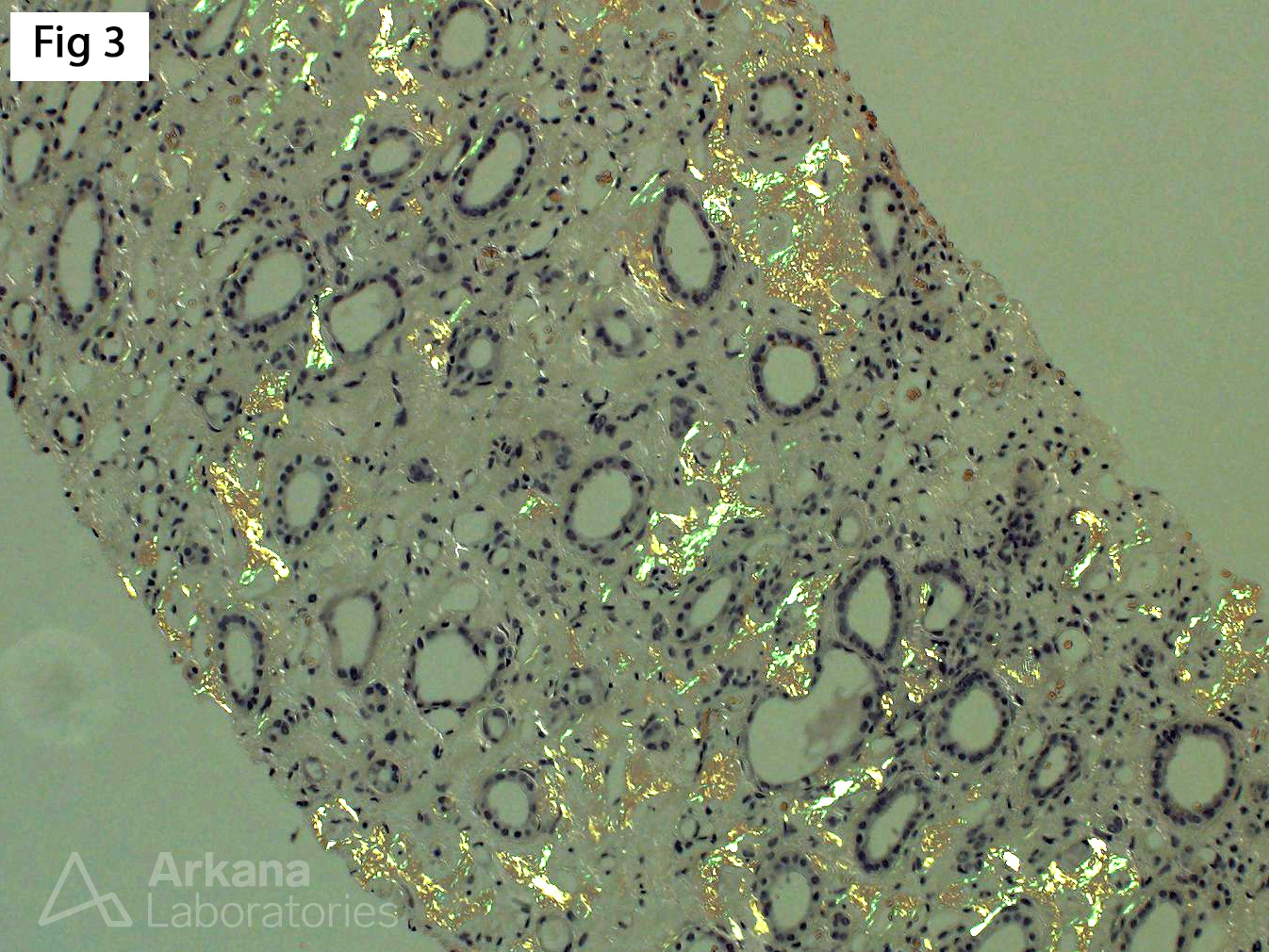
This renal biopsy from a 71-year-old female, who presented with progressively worsening renal failure, shows unremarkable glomeruli by light microscopy (Fig 1), with minimal associated tubulointerstitial chronic injury. Of note, multifocal PAS-pale, weakly argyrophilic, Congo red positive (Fig 2) deposits with apple green birefringence (Fig 3), consistent with amyloid deposits, are present exclusively within the interstitium of the renal medulla. No amyloid deposition is present within the renal cortex or involving vascular structures. The deposits show no light chain restriction by routine or paraffin immunofluorescence (not shown), making the possibility of light chain-type amyloidosis unlikely. Furthermore, an immunoperoxidase stain for AA amyloid is negative. The pattern of parenchymal involvement is highly suspicious for apolipoprotein A-IV amyloid. Confirmatory testing by mass spectrometry detected a peptide profile suggesting apolipoprotein A-IV amyloid deposition. Apolipoprotein A-IV is a rare form of amyloidosis which characteristically involves the interstitium of the renal medulla, with occasional peritubular accentuation. Interestingly, the renal cortex, glomeruli, and vessels are typically spared. Patients with this form of amyloidosis present with worsening renal function and absence of significant proteinuria.
Reference: Dasari et al. Clinical, biopsy, and mass spectrometry characteristics of renal apolipoprotein A-IV amyloidosis. Kidney Intl. 2016; 90(3): 658-664.
Quick note: This post is to be used for informational purposes only and does not constitute medical or health advice. Each person should consult their own doctor with respect to matters referenced. Arkana Laboratories assumes no liability for actions taken in reliance upon the information contained herein.