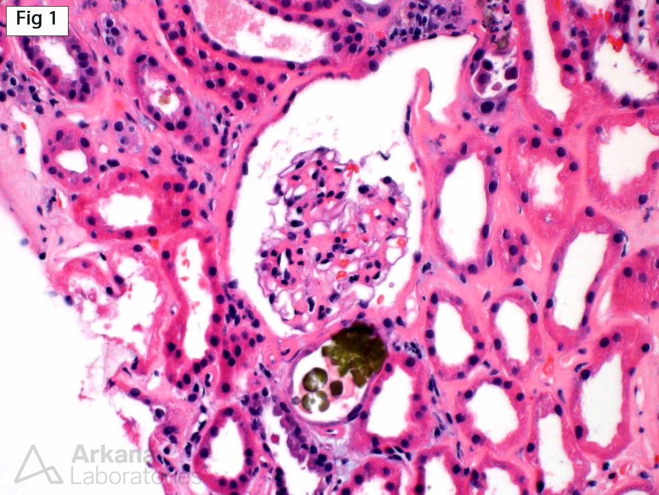
A renal biopsy was performed on this 75-year-old female who presented with acute liver failure, jaundice and acute renal failure (serum creatinine 6.4 mg/dl). The predominant biopsy findings are those of acute tubular injury with scattered translucent crystals which show a fan-like morphology (Fig 1) and birefringence upon polarization (Fig 2), consistent with calcium oxalate crystals. Due to the paucity of these crystals, they are favored to represent non-specific deposition in the setting of ongoing tubular injury. Interestingly, the deposited calcium oxalate crystals are bile-stained (Fig 1), a phenomenon which may be seen in the setting of hyperbilirubinemia. Although not present in this case, calcium phosphate deposits may also show green discoloration. It is important to recognize that these stained calcium deposits are not classified as bile casts, which have different morphologic and pathophysiologic characteristics (See reference).

Reference:
van Slambrouck CM, Salem F, Meehan SM, Chang A. Bile cast nephropathy is a common pathologic finding for kidney injury associated with severe liver dysfunction. Kidney Int. 2013; 84: 192-197.
Quick note: This post is to be used for informational purposes only and does not constitute medical or health advice. Each person should consult their own doctor with respect to matters referenced. Arkana Laboratories assumes no liability for actions taken in reliance upon the information contained herein.