Search & Filter All Posts
All Articles
(18 Results)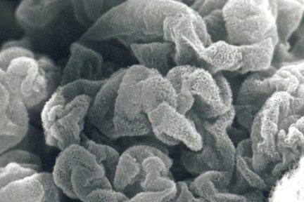
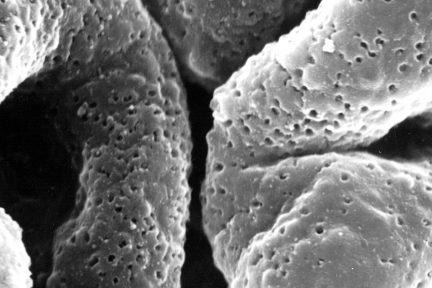
Membranous Glomerulonephritis
Todays eyeSCANdy image is of an acellular scanning EM of a glomerulus from a biopsy with membranous glomerulonephritis, stage II…
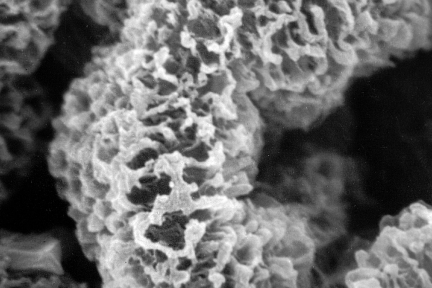
Membranous Glomerulonephritis Stage II
Todays acellular scanning EM from a biopsy with membranous glomerulonephritis, stage II shows a reticular pattern of the subepithelial GBM.…

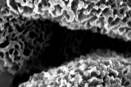
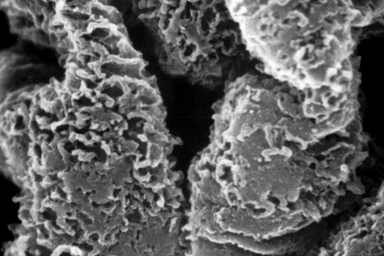
Glomerular Capillary Loops
This acellular scanning EM shows several glomerular capillary loops from a biopsy with membranous glomerulonephritis, stage II. The extracted deposits…
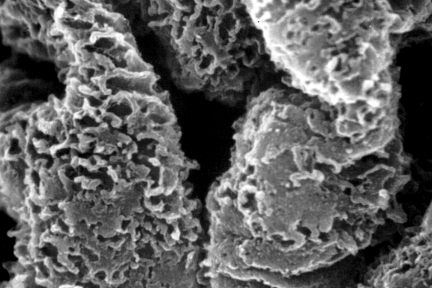
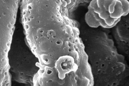
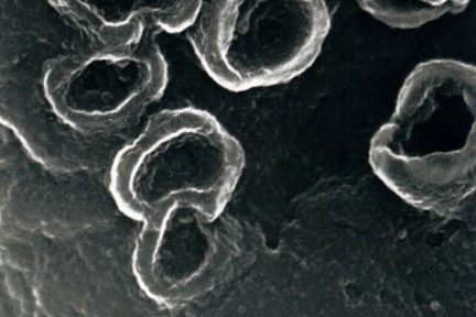
Depressions and Larger “Craters”
Todays acellular scanning EM from a biopsy with membranous glomerulonephritis shows depressions and larger “craters” along the subepithelial aspect of…
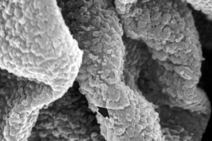
Subepithelial Immune Deposits Carpeting the GBM Surface
Todays eyeSCANdy image of an acellular scanning EM from a biopsy with membranous lupus nephritis shows subepithelial immune deposits (arrows,…

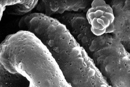
Extracted Immune Complexes
Todays acellular scanning EM from a biopsy with membranous glomerulonephritis, stage I shows numerous small shallow depressions representing the site…
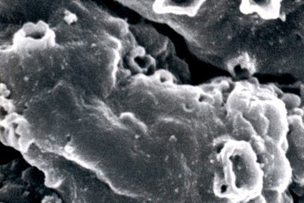
Membranous Glomerulonephritis Stage I
This acellular scanning EM from a biopsy with membranous glomerulonephritis, stage I shows numerous small shallow depressions representing the site…
Shallow Craters Along Subepithelial Aspect of the Basement Membrane
This acellular scanning EM from a biopsy with membranous glomerulonephritis, stage I shows numerous small shallow depressions and occasional larger…

Subepithelial Glomerular Basement Membrane
This acellular scanning EM from a biopsy with membranous glomerulonephritis, stage II shows a reticular appearance of the subepithelial glomerular…
Membranous Glomerulonephritis Stage III
This eyeSCANdy image shows acellular scanning EM from a biopsy with membranous glomerulonephritis, stage III. There is focal GBM bridging…
Membranous Glomerulonephritis Stage I
Todays eyeSCANdy image shows acellular scanning EM from a biopsy with membranous glomerulonephritis, stage I which shows numerous small shallow…
Membranous Glomerulonephritis
This eyeSCANdy image of acellular scanning EM shows craters at high power on the subepithelial aspect of the glomerular basement…
Glomerular Basement Membranes
This eyeSCANdy image shows an acellular scanning EM of glomerular basement membranes from a biopsy with membranous glomerulonephritis, stage II…
Craters on Glomerular Basement Membranes
Todays eyeSCANdyimage shows acellular scanning EM showing “craters” at high power on the subepithelial aspect of the glomerular basement membranes…
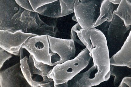
Basement Membrane Damage in Crescentic GN
Todays eyeSCANdy image shows acellular scanning EM showing GBMs with variably sized discrete perforations. This is the mildest form of…