Search & Filter All Posts
All Articles
(12 Results)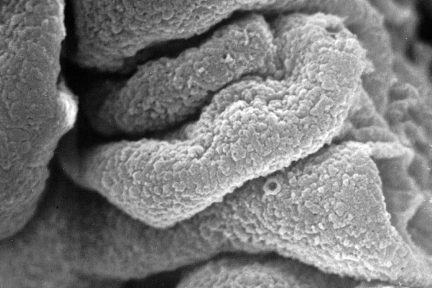
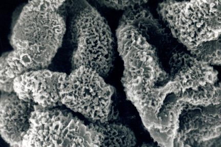
Subepithelial Immune Deposits Carpeting the GBM Surface
This acellular scanning EM from a biopsy with membranous lupus nephritis shows subepithelial immune deposits (not extracted) carpeting the GBM…
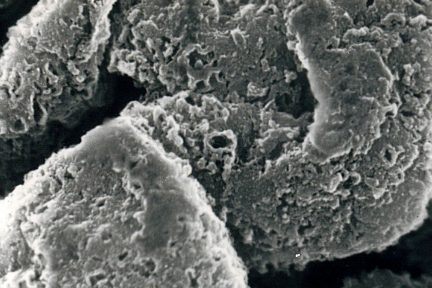
Subepithelial Immune Deposits Carpeting the GBM Surface
Todays acellular scanning EM from a biopsy with membranous lupus nephritis shows subepithelial immune deposits (not extracted) carpeting the GBM…
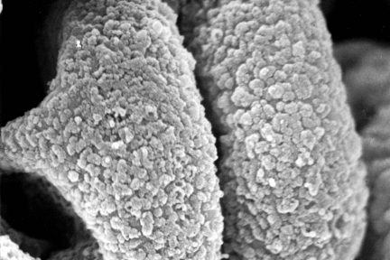
Glomerular Basement Membranes
This eyeSCANdy image shows an acellular scanning EM of glomerular basement membranes from a biopsy with membranous glomerulonephritis, stage II…
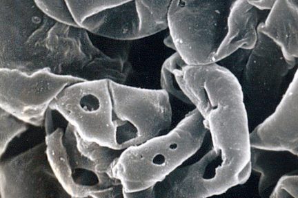
Basement Membrane Damage in Crescentic GN
Todays eyeSCANdy image shows acellular scanning EM showing GBMs with variably sized discrete perforations. This is the mildest form of…
Membranous Glomerulonephritis Stage II
This eyeSCANdy image shows acellular scanning EM from a biopsy with membranous glomerulonephritis, stage II. The extracted deposits previously resided…
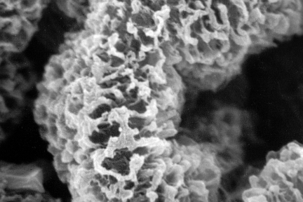
Stage III Membranous Glomerulonephritis
Todays eyeSCANdy shows acellular scanning EM from a biopsy with membranous glomerulonephritis, stage III! There is more extensive GBM bridging…
Diagnose This (January 19, 2021)
Based on the electron microscopic image, what would be your initial underlying clinical concern for the findings seen? …
Diagnose This (August 17, 2020)
What is your diagnosis for this patient with nephrotic syndrome? …
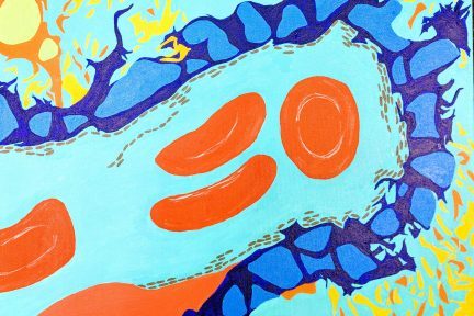
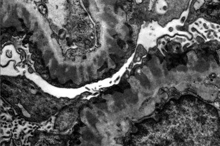
Art of Medicine: Membranous Glomerulopathy
The painting above depicts membranous glomerulopathy. A single glomerular capillary loop with confluent subepithelial and intramembranous electron dense deposits along…
KDIGO Connections: Membranous Glomerulopathy
Welcome to the first post in our new series KDIGO Connections, a series in which we are asking our nephrologist…
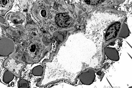
Resolving Membranous Glomerulopathy
A renal biopsy was performed on this 55-year-old female with a history of biopsy-proven membranous glomerulopathy status post immunosuppressive therapy,…
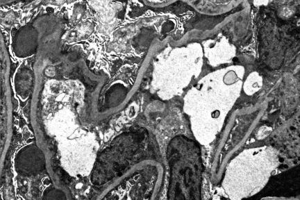
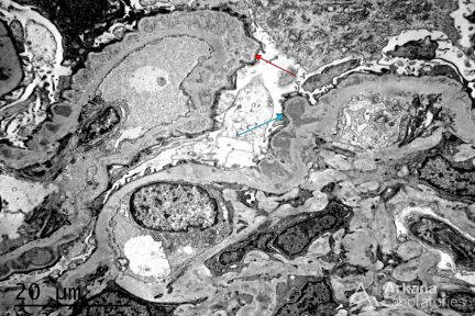
Subepithelial Humps
The depicted electron micrograph shows numerous, large and irregular subepithelial deposits which protrude from the glomerular basement membrane towards…