Search & Filter All Posts
All Articles
(1358 Results)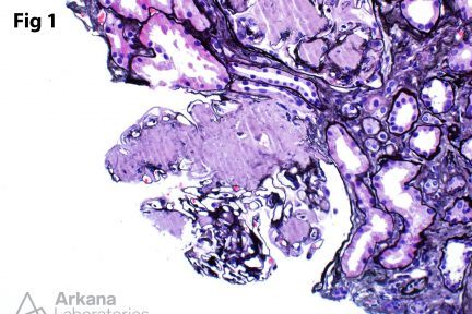
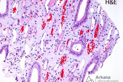
AA Amyloid
This biopsy from a patient with massive proteinuria showed diffuse and near-global involvement of glomeruli by amorphous deposits (Fig. 1)…

Wilson Corner
If you’ve ever met Dr. Jon Wilson, head of our neuropathology service, this set up outside his office makes perfect…
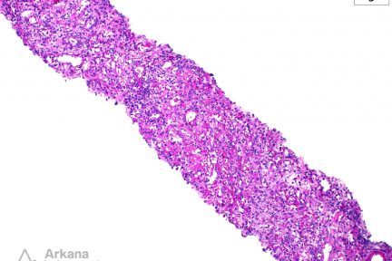
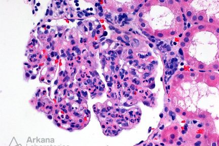
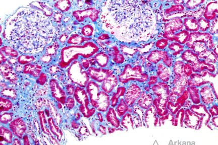
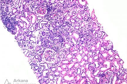
Megalocytic Interstitial Nephritis
This biopsy shows a chronic active interstitial nephritis with numerous interstitial foamy macrophages containing abundant PAS-positive cytoplasmic granules (Fig 1-3).…
Diagnose This! (August 14, 2017)
An immunofluorescence panel for IgA, IgG, IgM, C3, C1q, kappa and lambda was negative. What is your diagnosis? …
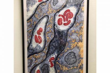
Arkana Artwork
We hang these two pieces proudly in one of our physician hallways. They were a gift from the talented nephropathologist…
Randall’s Plaques
Randalls plaques are microscopic calcium phosphate deposits in the basement membranes of the thin loops of Henle. These plaques act…
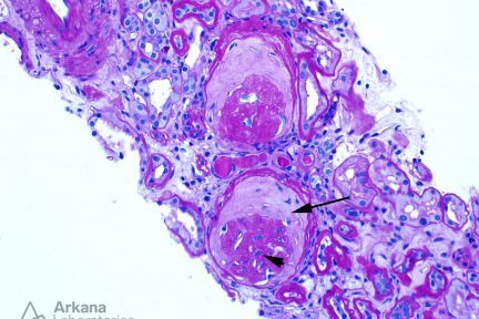
Global Glomerulosclerosis: Obsolescent Pattern
The image shows two globally sclerotic glomeruli exhibiting the so-called obsolescent pattern of glomerular sclerosis. The two key histologic features…
Pushing Glass (August 8, 2017)
A 69-year-old man with past medical history of hypertension presents with nephrotic syndrome and 7 gm/day proteinuria. Serological studies are…
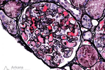
Fabrys
This biopsy shows glomeruli with visceral epithelial cells (podocytes) displaying ample vacuolated cytoplasm (Fig 1), which is highlighted on a…

Arkana Physician Spotlight: Dr. Kuperman
Dr. Michael Kuperman joined our staff in 2016, after previously practicing renal pathology for several years in Texas. He received…

Arkanines (August 4, 2017 )
Meet Butters! He enjoys picking fights with much larger dogs and stealing toys from his sister, but his cuteness makes…
Light Chain Deposition Disease and Light Chain Proximal Tubulopathy
An elderly but previously healthy patient presents with weakness and fatigue and was found to have a creatinine of 3.0…

Arkana Journal Club (August 3, 2017)
The Arkana Journal Club is happening right now. Follow @TheGoodMD on Twitter for live tweeting! #ArkanaJC http://jasn.asnjournals.org/content/28/7/2144 https://link.springer.com/article/10.1007%2Fs00467-016-3392-7
Tubulointerstitial Nephritis with Uveitis
Tubulointerstitial nephritis with uveitis (TINU) was first described as a distinct entity by Dobrin, et al. in 1975. TINU primarily…