An immunofluorescence panel for IgA, IgG, IgM, C3, C1q, kappa and lambda was negative. What is your diagnosis?
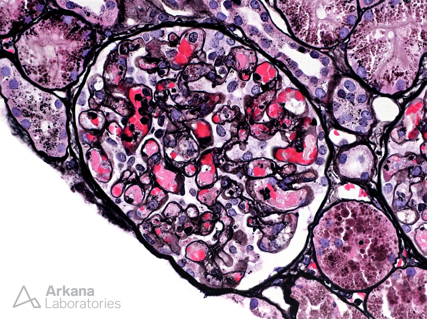
The photomicrograph shows a high power image of a glomerulus on Jones silver stain. The glomerulus displays mesangiolysis and enlarged capillary loops often filled with congested red blood cells and frequent fibrin thrombi. While a somewhat similar glomerular appearance can be seen in cryoglobulinemia, in the presence of a negative immunofluorescence panel these findings are most consistent with thrombotic microangiopathy.
Quick note: This post is to be used for informational purposes only and does not constitute medical or health advice. Each person should consult their own doctor with respect to matters referenced. Arkana Laboratories assumes no liability for actions taken in reliance upon the information contained herein.