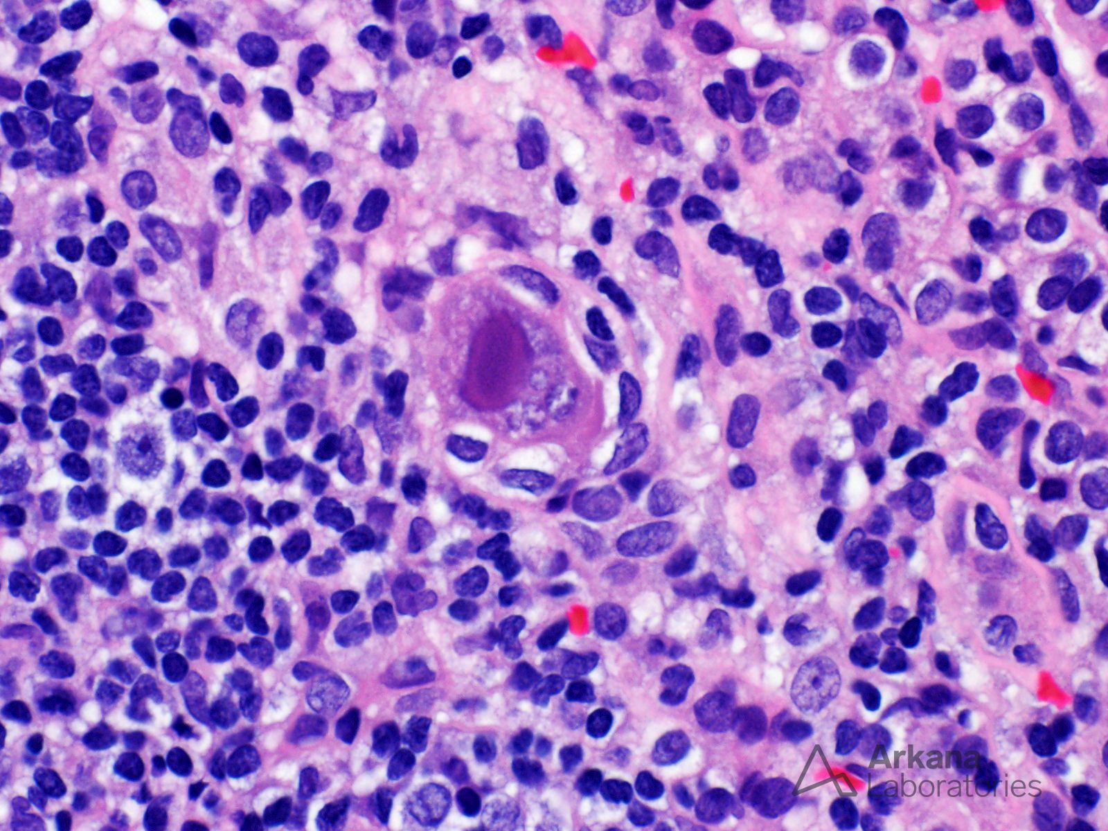
The biopsy from this adult kidney transplant recipient shows features of cytomegalovirus (CMV) infection, including prominent enlargement (“cytomegalo-”) of tubular epithelial cells and their nuclei, along with small basophilic inclusions which expand the cell cytoplasm. An immunostain for cytomegalovirus was also positive for viral antigen. In the kidneys, cytomegalovirus shows tropism for tubular epithelial and glomerular endothelial cells. The gold standard for the diagnosis of tissue-invasive CMV disease remains the histopathologic identification of the characteristic cytoplasmic and/or nuclear viral inclusions or positive immunohistochemical detection of cytomegalovirus viral antigens in tissue. Active cytomegalovirus disease may be donor-derived (iatrogenic) or it may represent primary infection or reactivation of latent cytomegalovirus infection in the recipient, and it still remains a cause of morbidity and graft loss in solid organ transplant recipients.
A summary of CMV management recommendations was published in 2013 by the Transplantation Society International CMV Consensus Group (Kotton et al. Updated international consensus guidelines on the management of cytomegalovirus in solid-organ transplantation. Transplantation. 2013 Aug 27;96(4):333-60). https://pubmed.ncbi.nlm.nih.gov/23896556/
Quick note: This post is to be used for informational purposes only and does not constitute medical or health advice. Each person should consult their own doctor with respect to matters referenced. Arkana Laboratories assumes no liability for actions taken in reliance upon the information contained herein.