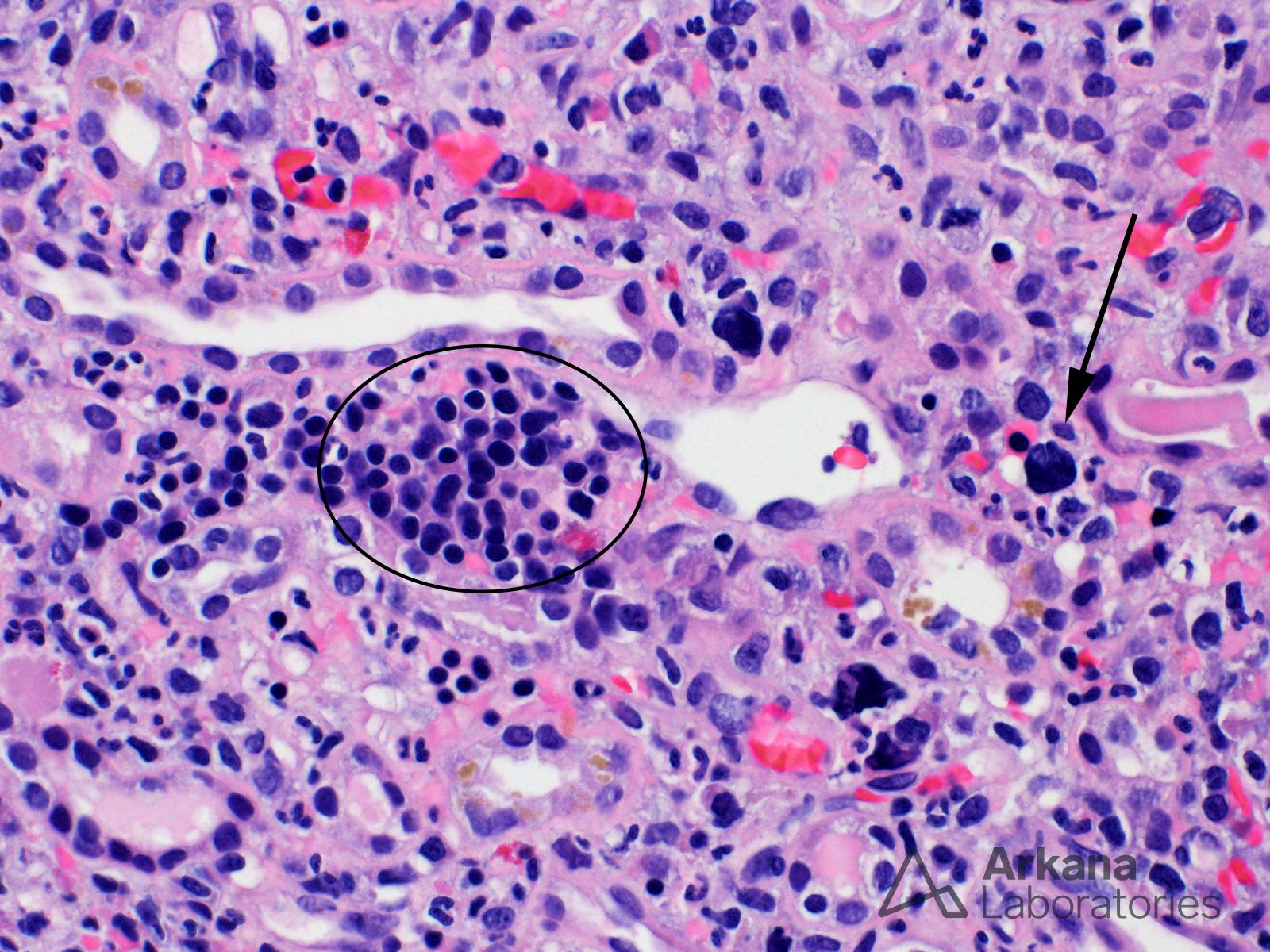
This renal biopsy shows extramedullary hematopoiesis (EMH), which is the presence of hematopoietic elements (erythroid, myeloid, and/or megakaryocytic) found outside of the bone marrow. The larger circle contains mostly erythroid precursors and the arrow identifies an immature megakaryocyte. Generally, the histologic differential diagnosis includes acute interstitial nephritis (erythroid precursors may be confused with mature inactive lymphocytes) and even some types of lymphoma (megakaryocytes may resemble atypical or malignant lymphoid cells). In difficult cases, lineage-specific immunohistochemical stains can be used to confirm the presence of hematopoietic cells.
Recognizing EMH is important because it often signifies impaired bone marrow function due to various conditions ranging from myelofibrosis to plasma cell myeloma. The 64-year-old man from whom this biopsy was taken presented with acute renal failure and pancytopenia. His kidney biopsy showed lambda light chain cast nephropathy (not shown) and a subsequent bone marrow biopsy showed extensive involvement by a lambda light chain-restricted plasma cell neoplasm.
Alexander MP, et al. Renal extramedullary hematopoiesis: interstitial and glomerular pathology. Mod Pathol. 2015 Dec;28(12):1574-83. PubMed:https://www.ncbi.nlm.nih.gov/pubmed/26449764
Quick note: This post is to be used for informational purposes only and does not constitute medical or health advice. Each person should consult their own doctor with respect to matters referenced. Arkana Laboratories assumes no liability for actions taken in reliance upon the information contained herein.