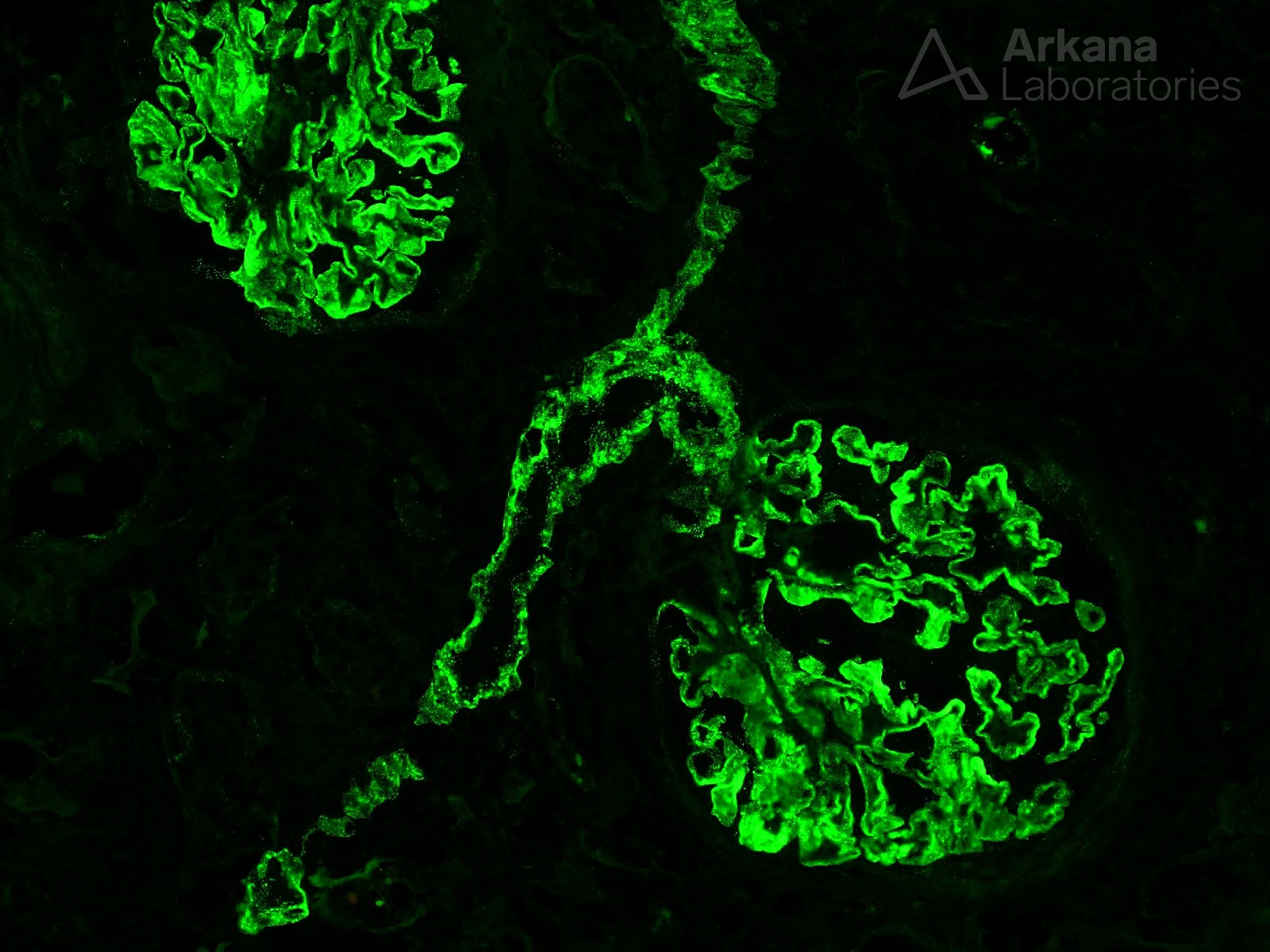
This immunofluorescence (IF) image shows extensive granular glomerular capillary wall IgG deposits, consistent with membranous glomerulopathy. The glomerular staining pattern as seen by IF is not specific for either primary or secondary membranous glomerulopathy. However, the prominent IgG deposits also involving the associated arteriole in this case are an important clue that helps argue for a secondary etiology. This patient, in fact, was found to have systemic lupus erythematosus.
Quick note: This post is to be used for informational purposes only and does not constitute medical or health advice. Each person should consult their own doctor with respect to matters referenced. Arkana Laboratories assumes no liability for actions taken in reliance upon the information contained herein.