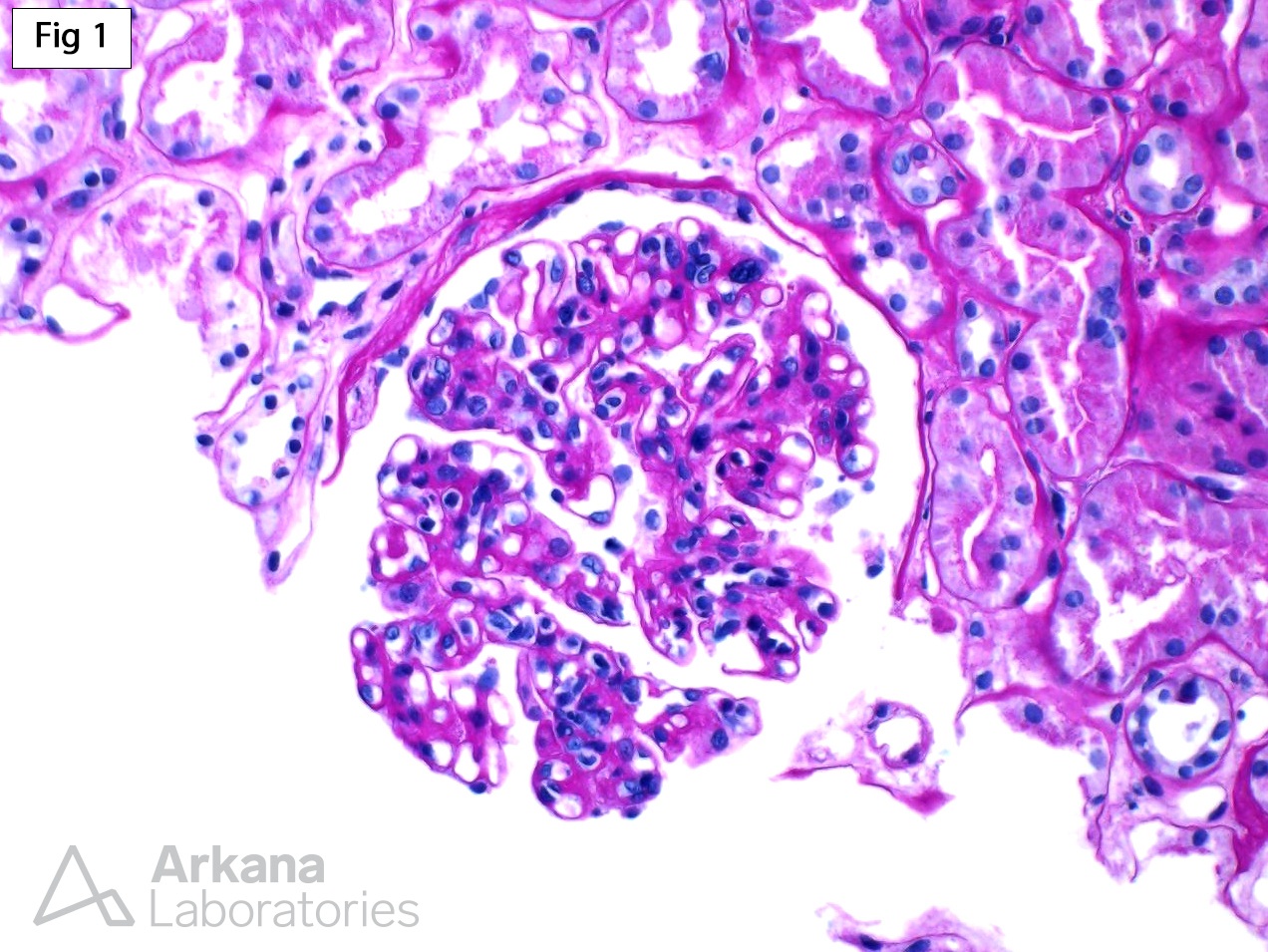
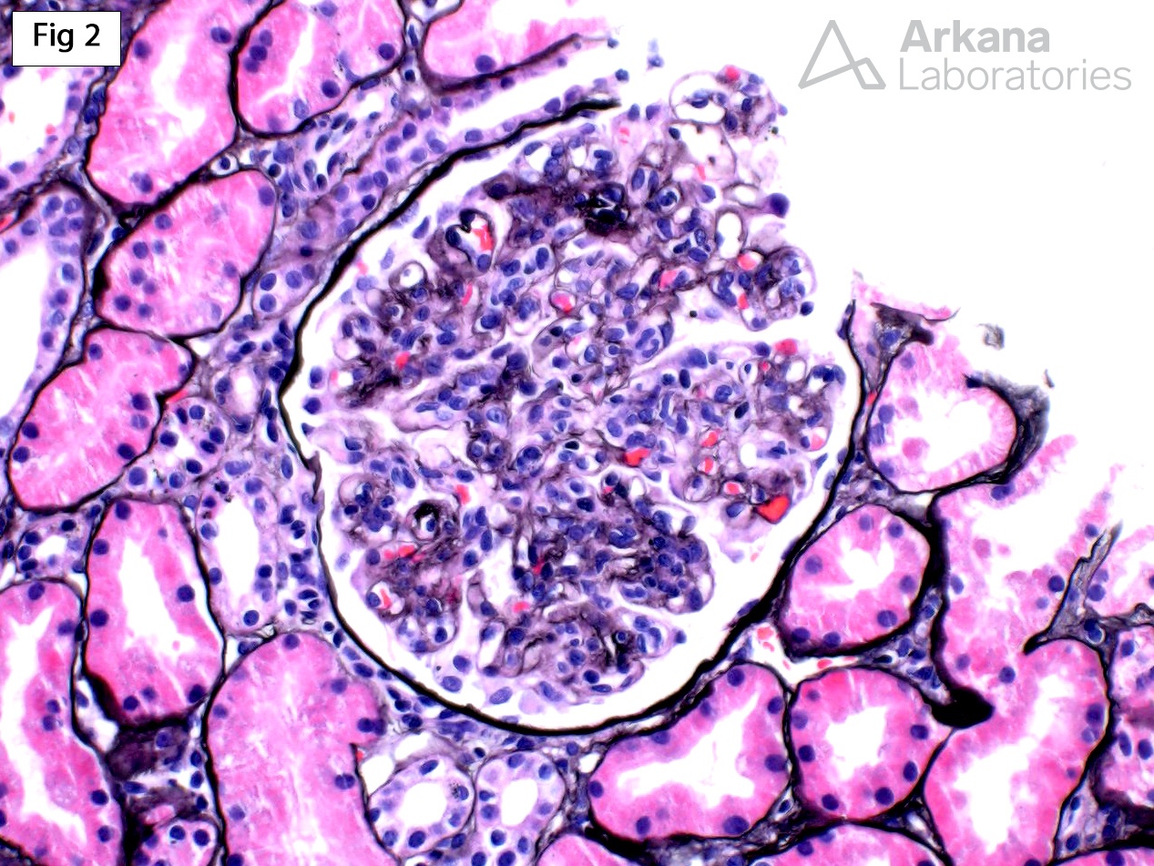
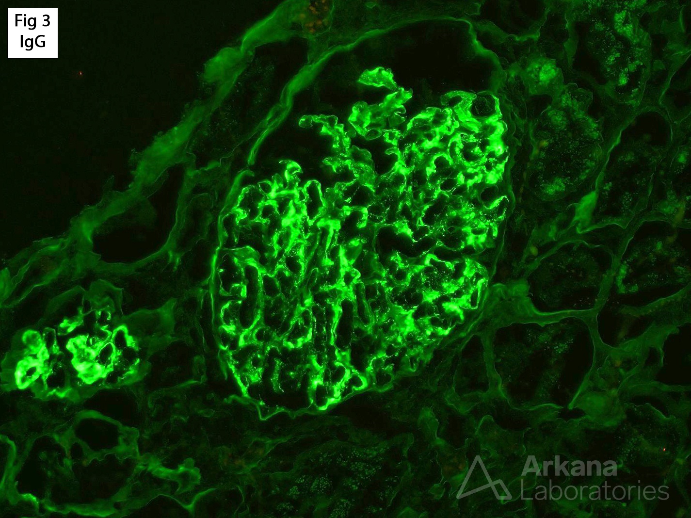
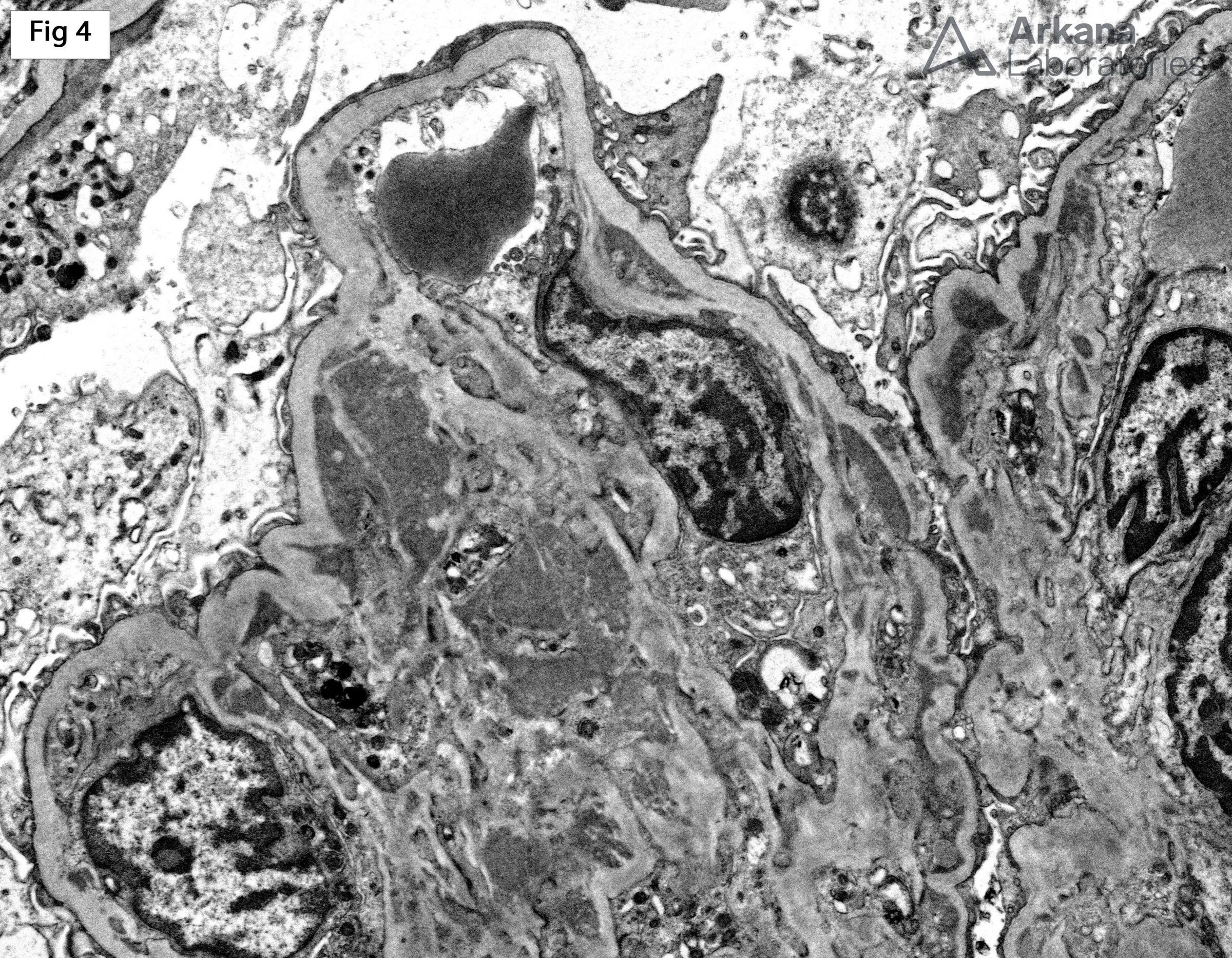
This biopsy was performed on an 81-year-male, status post renal transplant, who presented with increased creatinine (2.3 mg/dl) and microscopic hematuria. Light microscopic examination of the biopsy shows diffuse mesangial and mild endocapillary hypercellularity with rare double contour formation of the capillary loops (Fig 1 and 2). No significant tubulointerstitial inflammation, peritubular capillaritis or endothelialitis is present, to otherwise suggest and underlying component of acute T-cell or antibody mediated rejection. Immunofluorescence (Fig 3) shows mesangial and segmental capillary loop positive staining for IgG (3+), C3 (3+), C1q (1+) and lambda light chain (3+). All other stains, including kappa light chain are negative. Furthermore, the deposits are positive for IgG subclass 3 and negative for IgG1, IgG2 and IgG4. C4d is negative within peritubular capillaries. Lastly, electron microscopy shows mesangial and subendothelial deposits without definitive substructure (Fig 4). The overall biopsy findings are consistent with “proliferative glomerulonephritis with monoclonal IgG deposits”. This entity is characterized by an endocapillary proliferative, membranoproliferative or mesangial proliferative pattern of glomerular injury with monoclonal IgG deposits. The deposits are usually IgG3κ restricted (53.1%), but approximately 12.5% of cases show IgG3λ restriction (reference 1). Of note, although the deposits are composed of monoclonal immunoglobulins, the vast majority of patients do not appear to have an underlying hematologic malignancy; however, exclusion of such disorders, as well as cryoglobulinemia, is warranted. Similar to the case in discussion, recurrent and de novo forms of proliferative glomerulonephritis with monoclonal IgG deposits have been reported in renal allografts (reference 2 and 3).
References:
1) Nasr S, Satoskar A, Markowitz GS, et al. Proliferative Glomerulonephritis with Monoclonal IgG Deposits. J Am Soc Nephrol. 2009; 20: 2055-2064.
2) Nasr S, Sethi S, Cornell LD, et al. Proliferative glomerulonephritis with monoclonal IgG deposits recurs in the allograft. Clin J Am Soc Nephrol 2011; 6: 122-132
3) Albawardi A, Satoskar A, Von Visger J, et al. Proliferative glomerulonephritis with monoclonal IgG deposits recurs or may develop de novo in kidney allografts. 2011; 58: 276-281.
Quick note: This post is to be used for informational purposes only and does not constitute medical or health advice. Each person should consult their own doctor with respect to matters referenced. Arkana Laboratories assumes no liability for actions taken in reliance upon the information contained herein.