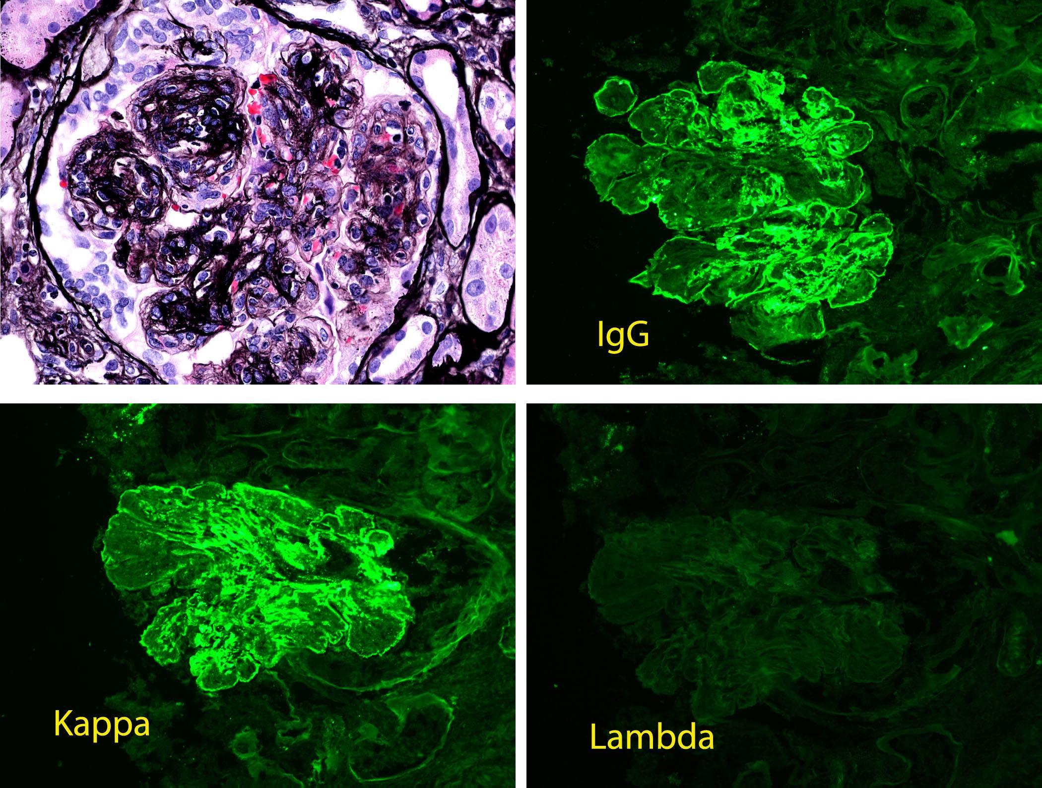
The glomerulus pictured for light microscopy shows membranoproliferative changes with mesangial hypercellularity, GBM duplication, and endocapillary proliferation. There is light chain-restricted staining for IgG kappa by immunofluorescence. The deposits were immune complex-type by electron microscopy (not pictured). The findings in this biopsy are consistent with the entity known as proliferative glomerulonephritis with monoclonal IgG deposits. A case series detailed the clinical findings in 37 patients with proliferative glomerulonephritis with monoclonal IgG deposits and found the underlying etiology was undetermined in the majority of cases. Most did not appear to be related to infection, autoimmune-related disease or hematologic malignancy (see reference below). Nevertheless, close inspection for an underlying lymphoproliferative disorder is warranted because approximately 30% of patients with these findings on biopsy have evidence of dysproteinemia. When this is the case, the lesion should be considered a monoclonal gammopathy of renal significance.
Reference: Nasr S, Satoskar A, et al. Proliferative Glomerulonephritis with Monoclonal IgG Deposits. J Am Soc Nephrol. 20:2055-2064, 2009.
Quick note: This post is to be used for informational purposes only and does not constitute medical or health advice. Each person should consult their own doctor with respect to matters referenced. Arkana Laboratories assumes no liability for actions taken in reliance upon the information contained herein.