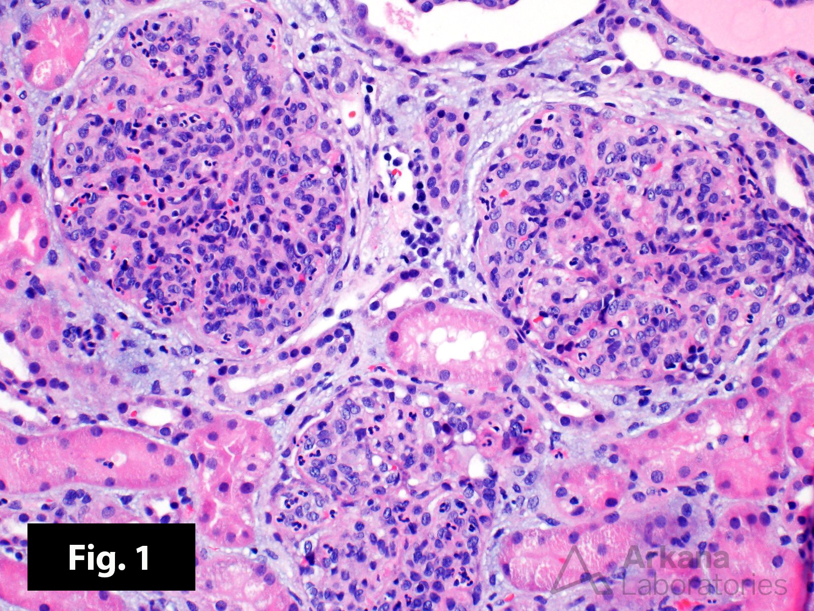
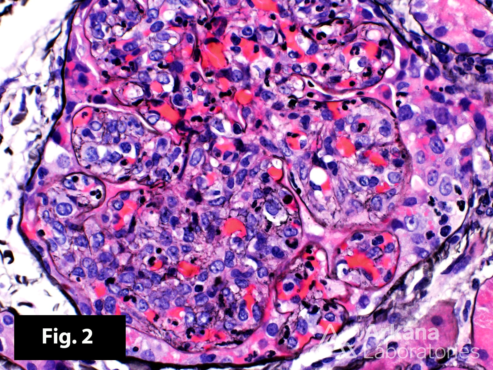
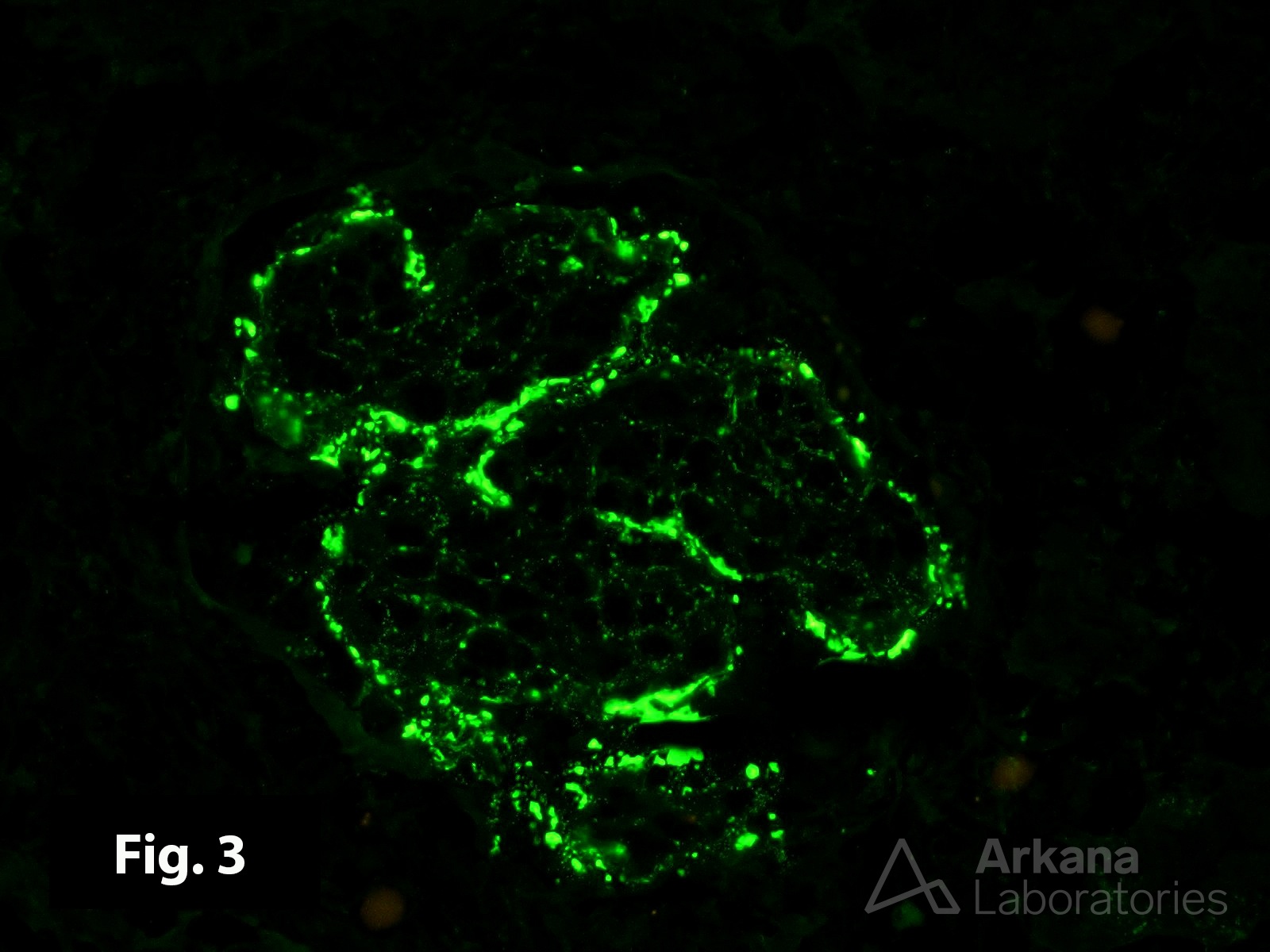
These renal biopsy images are from an 8-year-old boy who experienced the abrupt onset of hypertension, lower extremity edema, gross hematuria, and proteinuria about one week after seeing his pediatrician for a sore throat. The child had an elevated BUN and serum creatinine, and he was hypocomplementemic (C3). Figure 1 shows a diffuse proliferative (note the hypercellularity and closed capillary loops) and exudative (note the abundant neutrophils) glomerulonephritis. The Jones silver stain in Figure 2 confirms the presence of endocapillary, mesangial, and extracapillary hypercellularity. The immunofluorescence studies in Figure 3 show coarse, granular immune deposits along the peripheral capillary loops (anti-IgG is shown). Large subepithelial electron dense deposits were present by electron microscopy (not shown). The clinical history, laboratory results, and biopsy findings all support the diagnosis of an acute post-infectious glomerulonephritis. The patient likely had a preceding streptococcal infection.
Quick note: This post is to be used for informational purposes only and does not constitute medical or health advice. Each person should consult their own doctor with respect to matters referenced. Arkana Laboratories assumes no liability for actions taken in reliance upon the information contained herein.