The biopsy is from a 61-year-old man with a history of intermittent microscopic hematuria for many years who presents with recent 18-pound weight loss and nephrotic syndrome. His creatinine is mildly elevated at 1.3 mg/dL. He has 12.5 g of proteinuria and his serum albumin is 2.6 mg/dL.
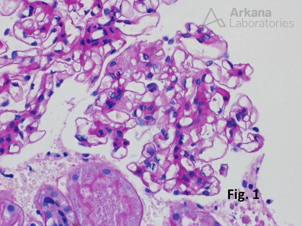
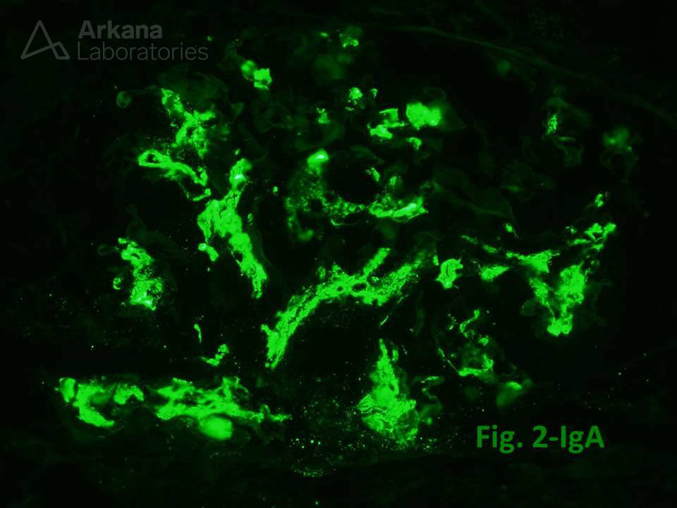
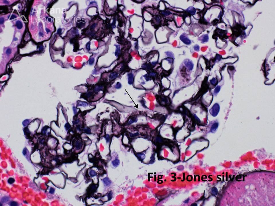
The biopsy shows diffuse mild mesangial matrix expansion with no necrosis or proliferative lesions (Fig. 1). Immunofluorescence microscopy shows extensive granular mesangial IgA deposits (3+) (Fig. 2), compatible with IgA nephropathy. Interestingly, the Jones methenamine silver stain also shows argyrophilic spikes involving capillary loops, which are most suggestive of spicular amyloid deposits (Fig. 3).
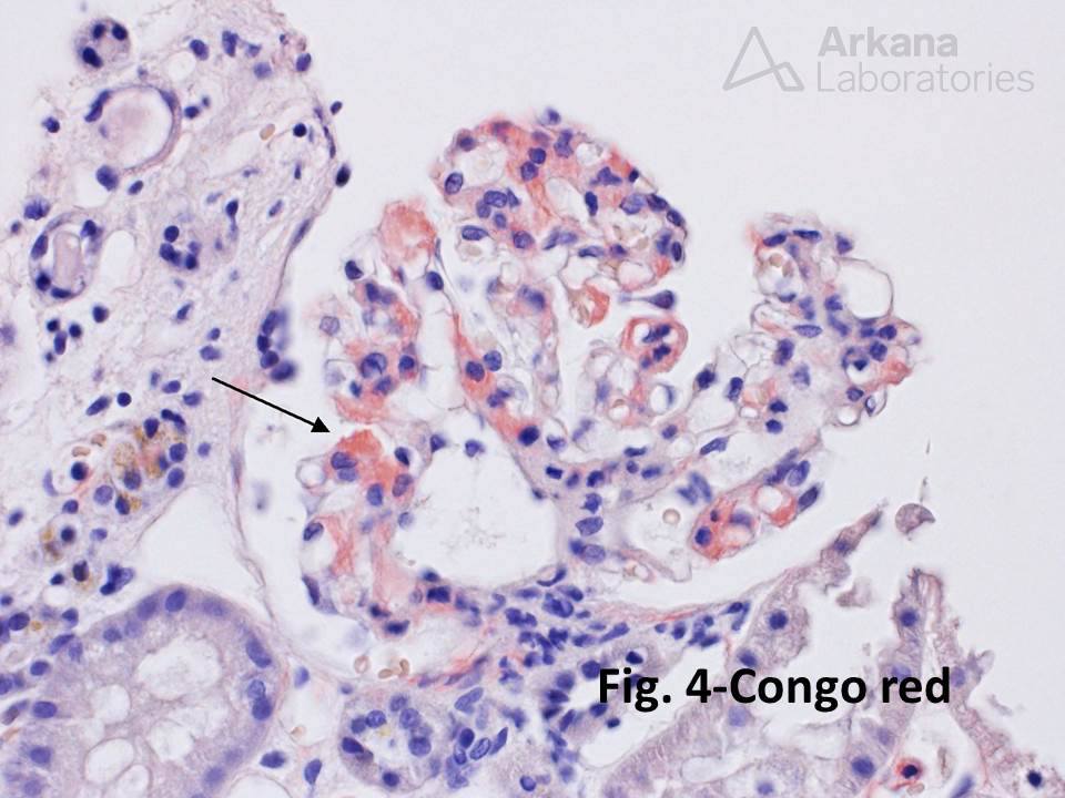
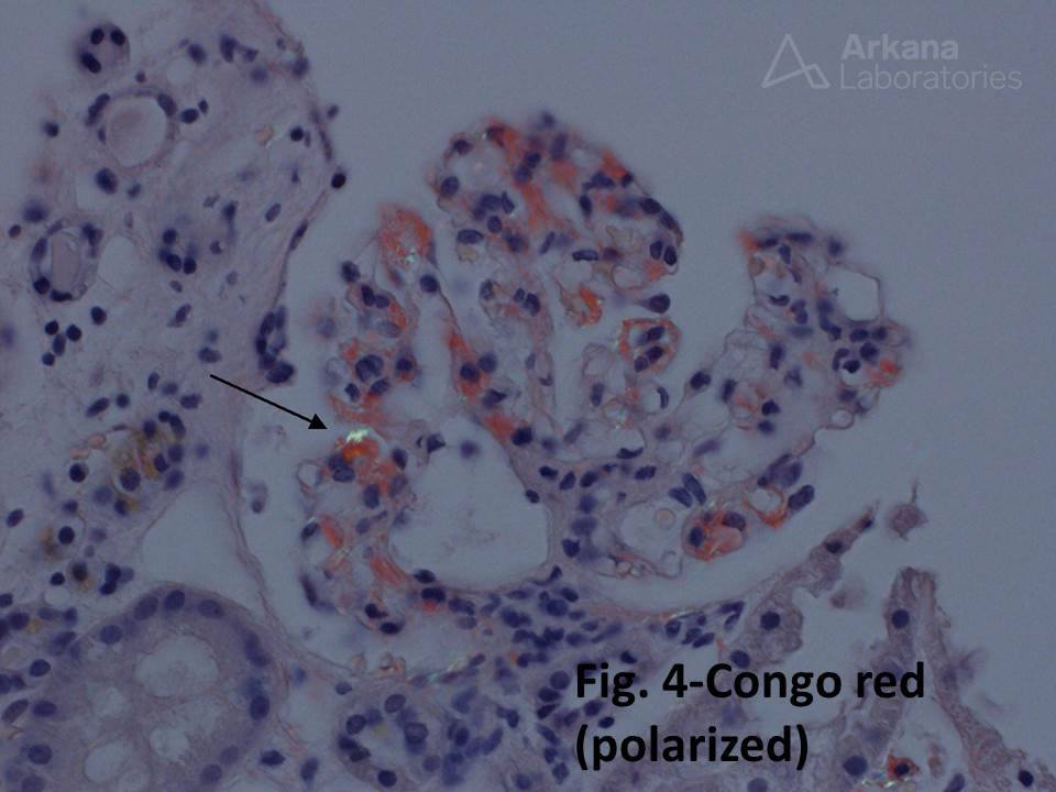
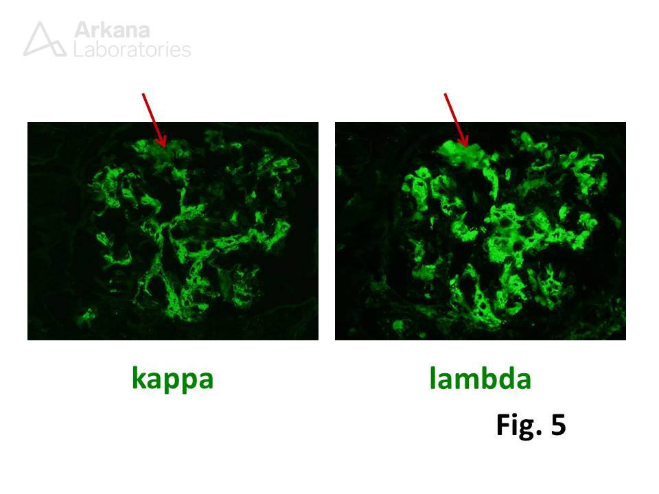
Congo red staining is positive for amyloid in these regions (Fig. 4), and immunofluorescence microscopy shows a smudgy 2+ reaction for lambda focally within the mesangium (Fig. 5). The corresponding areas on kappa staining are negative.
On clinical follow-up, the patient’s SPEP showed a monoclonal protein (lambda type), and serum free light chain testing showed a decreased K:L free light chain ratio.
Quick note: This post is to be used for informational purposes only and does not constitute medical or health advice. Each person should consult their own doctor with respect to matters referenced. Arkana Laboratories assumes no liability for actions taken in reliance upon the information contained herein.