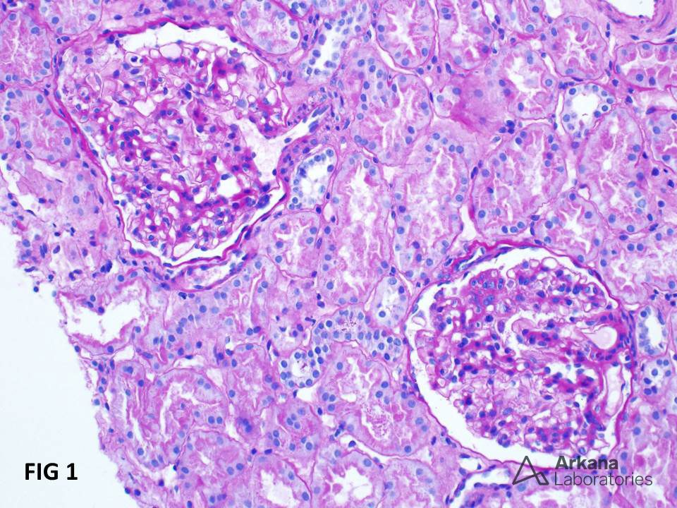
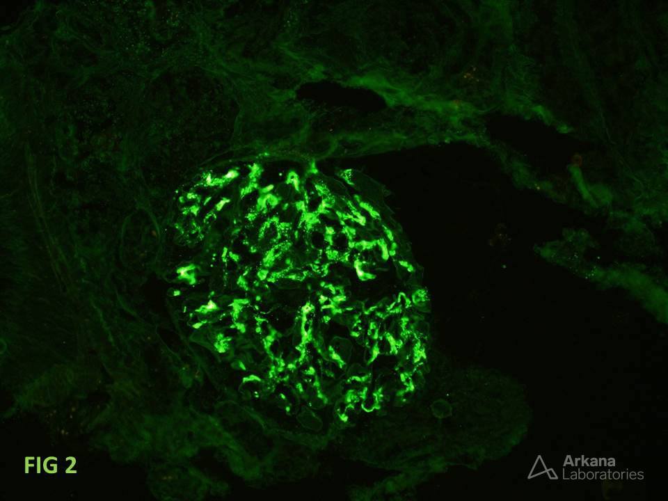
Figure 1 shows a renal biopsy from a 29-year-old man with no significant past medical history, who was found to have microscopic hematuria and non-nephrotic range proteinuria. The glomeruli show minimal mesangial matrix expansion and segmental hypercellularity. No crescents are identified. The surrounding tubules appear normal. Figure 2 shows dominant IgA mesangial deposits, consistent with IgA nephropathy. Remember that IgA deposits often persist and are seen in repeat biopsies even in patients who receive immunosuppressive therapy. Also, note the recent recommendation to include the presence or absence of crescents in the Oxford classification score (https://www.ncbi.nlm.nih.gov/pubmed/28341274).
Quick note: This post is to be used for informational purposes only and does not constitute medical or health advice. Each person should consult their own doctor with respect to matters referenced. Arkana Laboratories assumes no liability for actions taken in reliance upon the information contained herein.